. 2010 Apr;49(4):337-52.
doi: 10.1002/mc.20603.

Vitamin D receptor ligands, adenomatous polyposis coli, and the vitamin D receptor FokI polymorphism collectively modulate beta-catenin activity in colon cancer cells

Affiliations

Vitamin D receptor ligands, adenomatous polyposis coli, and the vitamin D receptor FokI polymorphism collectively modulate beta-catenin activity in colon cancer cells

Jan B Egan et al. Mol Carcinog. 2010 Apr.

Abstract

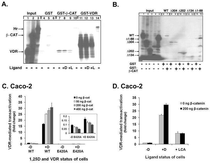
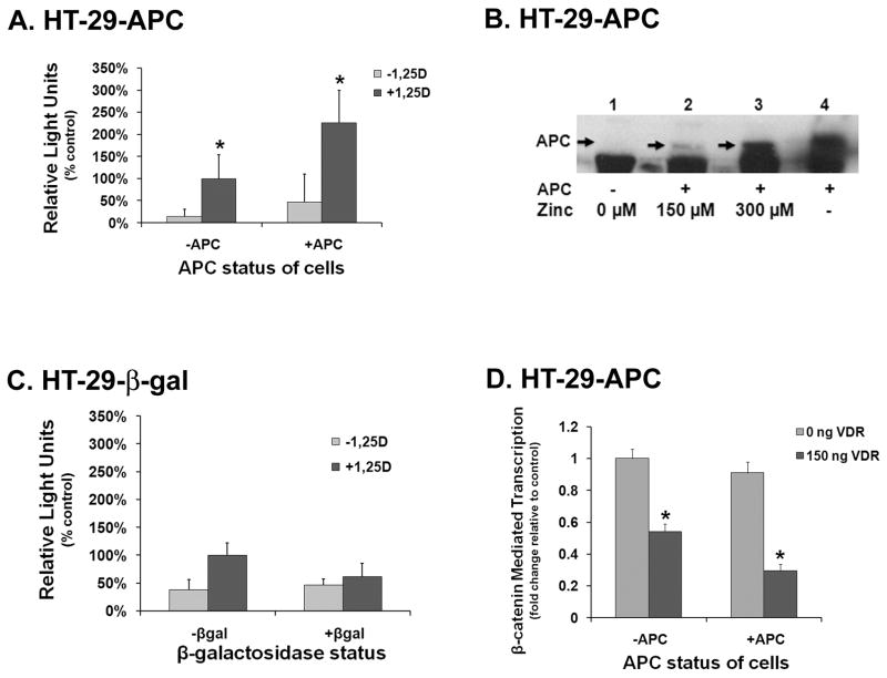
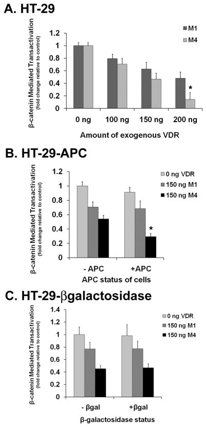
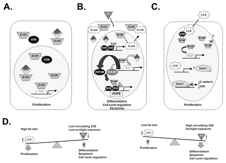
The activity of beta-catenin, commonly dysregulated in human colon cancers, is inhibited by the vitamin D receptor (VDR), and this mechanism is postulated to explain the putative anti-cancer activity of vitamin D metabolites in the colon. We investigated the effect of a common FokI restriction site polymorphism (F/f) in the human VDR gene as well as the effect of anti-tumorigenic 1,25-dihydroxyvitamin D(3) (1,25D) and pro-tumorigenic lithocholic acid (LCA) VDR ligands on beta-catenin transcriptional activity. Furthermore, the influence of a major regulatory protein of beta-catenin, the APC tumor suppressor gene, on VDR-dependent inhibition of beta-catenin activity was examined. We report herein that beta-catenin-mediated transcription is most effectively suppressed by the VDR FokI variant F/M4 when 1,25D is limiting. Using Caco-2 colorectal cancer (CRC) cells, it was observed that VDR ligands, 1,25D and LCA, both suppress beta-catenin transcriptional activity, though 1,25D exhibited significantly greater inhibition. Moreover, 1,25D, but not LCA, suppressed endogenous expression of the beta-catenin target gene DKK-4 independent of VDR DNA-binding activity. These results support beta-catenin sequestration away from endogenous gene targets by 1,25D-VDR. This activity is most efficiently mediated by the FokI gene variant F/M4, a VDR allele previously associated with protection against CRC. Interestingly, we found the inhibition of beta-catenin activity by 1,25D-VDR was significantly enhanced by wild-type APC. These results reveal a previously unrecognized role for 1,25D-VDR in APC/beta-catenin cross talk. Collectively, these findings strengthen evidence favoring a direct effect on the Wnt-signaling molecule beta-catenin as one anti-cancer target of 1,25D-VDR action in the colorectum.


Figures

Figure 1
Figure 1. Probing of protein-protein interactions between β-catenin and human VDR
A. GST fusion proteins were used to assess binding of radiolabeled bait and glutathione-immobilized prey proteins. Human VDR, β-catenin (β-CAT) or rat hairless (Hr) expression plasmids (1.0 μg) containing the T7 promoter were used as a template in an in vitro transcription/translation (IVTT) reaction to generate 35S-methionine-labeled bait proteins. VDR-containing lysates were also incubated with 10−6 M 1,25D (+D), 10−4 M lithocholic acid (+L), or ethanol vehicle (−) as indicated below the lanes 7–9. The lysates were then combined with either 20 μL of glutathione-S-transferase (GST) fusion protein alone bound to Sepharose beads (GST; lanes 4–6), GST-WT-VDR (lanes 7–10) or GST-β-catenin-S37A (lanes 11–14) for 30 min. GST-VDR beads were pre-incubated with the indicated ligands at the same concentration employed for the lysates (lanes 11–13). The beads were then washed extensively and the amount of coprecipitated VDR, β-catenin, or hairless was detected by electrophoresis of denatured bead samples followed by autoradiography. The arrows indicate the migration position of VDR, β-catenin and hairless. Aliquots (5%) of all radiolabeled protein inputs are shown at far left (lanes 1–3). The autoradiography gel is representative of 4 independent experiments. B. VDR truncation mutants define one region of the receptor required for contact with human β-catenin. WT and C-terminally truncated VDR proteins were generated in the IVTT system and incubated with either 20 μL of GST-β-CAT-Sepharose or GST-Sepharose only (as indicated below each lane) for 30 min. The beads were then washed and analyzed as described in the legend to Fig. 1A. Three C-terminal truncated receptor mutants are shown, with a stop codon inserted at the indicated residue position in VDR (Δ304, Δ202, Δ134). An N-terminal truncated receptor that is missing the first 88 amino acids was also employed (Δ1–88). The autoradiography gel is representative of 3 independent experiments. C. WT and E420A mutated VDR was transfected into Caco-2 cells along with the indicated amount of β-catenin, human CYP24A1 promoter-luciferase reporter and renilla plasmid. Cells were treated with ethanol vehicle or 10−8 M 1,25D prior to luciferase assay. VDR-mediated transactivation was measured as firefly luciferase output. Firefly values were normalized for transfection efficiency (using renilla luciferase) and expressed as the ratio of Firefly/Renilla relative light units (RLUs) prior to calculation of fold effects. The data illustrated are representative of 3 independent experiments with triplicate samples in each group. Error bars indicate standard deviation. When the change in activity from 0 to 400 ng of β-catenin was compared with a t-test for the WT VDR −1,25D and +1,25D groups, the increased activity was statistically significantly greater for the +1,25D group (P = 0.002), and regression modeling revealed a statistically significant dose-dependent increase in activity with increased β-catenin (P-trend = 0.001) for the wildtype VDR in the presence of ligand. A Student’s t-test and ANOVA indicated that the E420A VDR exhibited less than 1% of the activity of WT VDR, both in the presence (P = 0.0001) and absence (P = 0.003) of 1,25D; differences remained statistically significant after employing Tukey’s HSD. No increase in activity for E420A was observed with the addition of β-catenin (P = 0.22 with ANOVA). D. WT VDR was transfected into Caco-2 cells along with the indicated amount of β-catenin, human CYP24A1 promoter-luciferase reporter and renilla plasmid. Cells were treated with ethanol vehicle, 10−7 M 1,25D, or 70 μM LCA prior to luciferase assay. VDR-mediated transactivation was measured as firefly luciferase output, and then normalized as in (C). The data shown are representative of 3 independent experiments with triplicate samples in each group. Error bars indicate standard deviation. As evaluated with ANOVA, 1,25D and LCA both stimulated transactivation (P = 0.0001 for both ligands); results remained significant after adjustment with Tukey’s HSD. Transfection of β-catenin further augmented transcription by 1,25D-VDR (P = 0.0004), but not by LCA-VDR (P = 0.93); Student’s t-tests.
Figure 2
Figure 2. Effect of 1,25D and LCA on the interaction of VDR and β-catenin, transcriptional activity of β-catenin and endogenous DKK-4 expression
A. Representative graph of mammalian two hybrid assay utilizing Caco-2 cells to assess the effect of 1,25D and LCA on the interaction of VDR and β-catenin. Caco-2 cells were transfected with VDR prey, β-catenin bait, and then treated with vehicle (−1,25D), 10−8 M 1,25D or 70 μM LCA. Cells were then lysed and subjected to luciferase assay. VDR and β-catenin interaction was measured as firefly luciferase output (RLUs). Error bars indicate standard deviation. In the presence of 1,25D, interaction between VDR and β-catenin was increased 12-fold, while LCA increased the association by 8-fold (P-value for ANOVA = 0.0001; after Tukey’s HSD all pairwise comparisons remained statistically significant at a 99% confidence level). B. TOPFlash assay measuring the effect of VDR on β-catenin transcriptional activity. HT29 cells were transfected the indicated amounts of exogenous F/M4 VDR, as well as human β-catenin and the TOPFlash vector. Cells were treated with vehicle −1,25D) or 10−9 M 1,25D for 24 hours prior to cell lysis and luciferase assay. Transcriptional activity was measured as firefly luciferase light output. Error bars indicate standard deviation. Using regression analyses, a statistically significant dose-response trend for suppression of beta-catenin activity with increasing VDR in the presence of ligand was observed (P-trend = 0.0001). C. Representative graph of the TOPFlash assay in Caco-2 cells transfected with F/M4 VDR, TOPFlash vector, and β-catenin. Cells were treated with vehicle (−1,25D), 10−8 M 1,25D or 70 μM LCA and β-catenin transcriptional activity was measured as firefly luciferase output. Error bars indicate standard deviation. ANOVA reveals suppression of β-catenin activity at 25 ng VDR by 1,25-D but not LCA (* P = 0.04; though pairwise comparisons are not statistically significant after employing Tukey’s HSD). At 150 ng VDR, LCA was less effective at inhibiting β-catenin transactivation as compared to 1,25D (30%; ** P = 0.019 and 97%; P = 0.001), respectively; pairwise comparisons remained significant after Tukey’s HSD. D. Quantitative RT-PCR to evaluate the effect of 1,25D and LCA on DKK-4 expression in Caco-2. Cells were transfected with 0 ng, 600 ng wild-type VDR, or 600 ng DNA-binding mutant VDR, and treated with either vehicle or 10−7 M 1,25D (bars left of dashed line), or with vehicle, 10−7 M 1,25D, or 70 μM LCA (bars right of dashed line) 24 hours prior to RNA extraction. PCR was performed using the Applied Biosystems 7500 Fast. Expression was normalized to GAPDH endogenous control. Fold change was determined by comparing ligand treated samples to the vehicle treated sample for that transfection group. The reference group is assigned a value of one. All statistical analyses were student’s t-tests. * P = 0.001 when comparing the −1,25D and +1,25D groups that did not receive exogenous VDR. Cells with mutant and wildtype VDR demonstrated similar DKK-4 expression (P = 0.0004 for both M4 and mutant VDR). Data in all panels are representative of 3 independent experiments with triplicate samples in each group.
Figure 3
Figure 3. APC enhances both the interaction of VDR and β-catenin as well as the VDR-mediated suppression of β-catenin transcriptional activity
A. A mammalian two hybrid assay was utilized in HT-29-APC inducible cells to assess if VDR and β-catenin interact within an intact cell in the presence and absence of full length APC. HT-29-APC cells were transfected with VDR prey, β-catenin bait, and renilla plasmids and then treated with vehicle or 10−8 M 1,25D as well as vehicle or 150 μM zinc chloride (to induce full length APC). Cells were then lysed and subjected to luciferase assay. Interaction was measured as firefly luciferase output. Firefly output numbers were normalized using a firefly/renilla ratio to control for transcription efficiency. Percent change in the firefly/renilla ratio was determined by comparing all groups to the −APC(−Zn)/+1,25D group. This reference group is assigned a value of 100%. Error bars indicate standard deviation. * P < 0.001 when comparing the −APC and +APC groups which received 1,25D. B. Western blot of HT-29-APC cell lysates treated with zinc. HT-29-APC cells were treated with vehicle, 150 μM zinc chloride or 300 μM zinc chloride (lanes 1, 2 and 3, respectively) and allowed to incubate overnight prior to lysis. Lysates were then subjected to western blotting, using a chemiluminescence detection system and then exposed to film. HCT-116 cell lysates were used as an APC positive control (lane 4). C. Mammalian two hybrid assay in HT-29-βgalactosidase cells. HT-29-βgalactosidase cells were transfected with VDR prey, β-catenin bait, and renilla plasmids and then treated with vehicle or 10−8 M 1,25D as well as vehicle or 150 μM zinc chloride (to induce β-galactosidase). Cells were then lysed and subjected to luciferase assay. Interaction was measured as firefly light output. Firefly light output numbers were normalized using a firefly/renilla ratio to control for transfection efficiency. Percent change in the firefly/renilla ratio was determined by comparing all groups to the −βgalactosidase (−Zn)/+1,25D reference group which is assigned a value of 100%. Error bars indicate standard deviation. D. TOPFlash assay in HT-29-APC cells to assess the functional effects of full length APC on β-catenin transcriptional activity. F/M4 VDR was transfected into HT-29-APC cells along with a TOPFlash vector and renilla plasmid. Cells were treated with ethanol vehicle or 10−9 M 1,25D as well as vehicle or 150 μM zinc chloride prior to lysing and luciferase assay. Transcriptional activity was measured as firefly luciferase light output and the firefly values normalized by calculating fold effects via a +1,25D/−1,25D ratio. Fold transcription was determined by comparing the +1,25D/−1,25D ratio of each VDR group to the reference group of 0 ng VDR and no full length APC. The reference group is assigned a value of one. Error bars indicate standard deviation. * P < 0.001 when comparing the APC and +APC groups which received 1,25D. The data in all panels (except B) are the mean of three independent experiments with triplicate samples in each group.
Figure 4
Figure 4. Differential effect of FokI isoforms and APC on β-catenin transcriptional activity
A. Varying amounts of exogenous f/M1 and F/M4 VDR were transfected into HT-29 parental cells along with a TOPFlash vector and renilla plasmids. Cells were treated with ethanol vehicle or 10−9 M 1,25D prior to lysing and luciferase assay. Transcriptional activity was measured as luciferase light output and normalized by utilizing a +1,25D/−1,25D ratio of the firefly output. Fold transcription was determined by comparing the +1,25D/−1,25D firefly ratio of each VDR concentration to the reference group of 0 ng VDR for each isoform of VDR. The two reference groups are assigned a value of one. Error bars indicate standard deviation. Regression models revealed statistically significant trends (P = 0.0001) for both variants with increasing doses of VDR. A Student’s t-test indicated that the suppression of the TCF/LEF reporter was significantly greater for the M4 variant as compared to the M1 variant (* P = 0.0001). B. 150 ng of f/M1 and F/M4 VDR were transfected into HT-29-APC cells with a TOPFlash vector and renilla plasmid. Cells were treated with vehicle, 150 μM zinc, and/or 10−9 M 1,25D prior to lysing and luciferase assay. Zinc was used to induce the expression of APC (see Fig. 3). Transcriptional activity was measured as luciferase light output. The firefly output was normalized as in (A) and fold transcription determined by comparing the +1,25D/−1,25D ratio of each VDR group to the reference group of 0 ng VDR and no full length APC. The reference group is assigned a value of one. Error bars indicate standard deviation. P = 0.45 for the comparison of −APC vs. +APC in the M1 group (Student’s t-test). * P < 0.0001 for the comparison of −APC vs. +APC in the M4 group (Student’s t-test). All pairwise comparisons remained significant at the 99% confidence level after Tukey’s HSD. C. 150 ng exogenous f/M1 and F/M4 VDR were transfected into HT-29-βgalactosidase cells with a TOPFlash vector and renilla plasmid. Treatments and transcriptional activity was determined as in (B). Using a student’s t-test, no changes in β-catenin transcriptional activity was observed with the addition of β-gal in either the M1 (P = 0.97) or M4 (P = 0.73) variant. The data in all panels are the mean of three independent experiments with triplicate samples in each group.
Figure 5
Figure 5. Proposed model of VDR-β-catenin interactions
A. Absence of 1,25D. There is weak interaction (indicated by single line) of VDR, β-catenin and APC. β-catenin is able to freely translocate to the nucleus and bind to the promoter of target genes resulting in cell proliferation. B. Presence of 1,25D (D3). There is a robust interaction (indicated by four lines) of 1,25D-VDR and β-catenin (bottom), with stronger binding/sequestration by the F/M4 VDR isoform (center). The β-catenin in this complex is unable to activate TCF/LEF-driven proliferation genes, and instead is diverted towards VDRE-mediated transcription of 1,25D target genes regulating differentiation, cell cycle regulation and apoptosis. The 1,25D-VDR-RXR complex (with or without β-catenin) also binds to the VDRE in the promoter region of the E-cadherin gene to upregulate this protein which then facilitates the movement of β-catenin from the nucleus to the plasma membrane (top). C. Presence of lithocholic acid. LCA-VDR does not interact as robustly (indicated by two lines) with β-catenin compared to 1,25D-VDR. Thus, in the presence of an increased LCA/1,25D ratio, there is a relative elevation in the free pool of β-catenin resulting in more TCF/LEF-mediated expression of proliferative genes (top). LCA also upregulates the Snail1 transcriptional repressor (center) which has been previously shown to inhibit the expression of E-cadherin and VDR (bottom) and to prevent the nuclear exit of β-catenin, further increasing the pool of nuclear β-catenin and shifting the balance towards upregulation of β-catenin proliferative target genes. D. The role of dietary and environmental factors. A high fat diet, which results in the production of elevated colonic LCA, and/or low circulating 25D and low sunlight exposure shifts the “equilibrium” towards proliferation over differentiation. Conversely, high circulating 25D and adequate sunlight exposure, coupled with a low-fat diet, will lead to colonocytes that support the processes of differentiation, apoptosis and cell cycle regulation.

Similar articles

Cited by

References

    1. Mangelsdorf DJ, Thummel C, Beato M, et al. The nuclear receptor superfamily: the second decade. Cell. 1995;83(6):835–839. - PMC - PubMed
    1. Wood RJ, Fleet JC. The genetics of osteoporosis: vitamin D receptor polymorphisms. Annu Rev Nutr. 1998;18:233–258. - PubMed
    1. Jurutka PW, Remus LS, Whitfield GK, et al. The polymorphic N terminus in human vitamin D receptor isoforms influences transcriptional activity by modulating interaction with transcription factor IIB. Mol Endocrinol. 2000;14(3):401–420. - PubMed
    1. Fang Y, van Meurs JB, d’Alesio A, et al. Promoter and 3′-untranslated-region haplotypes in the vitamin d receptor gene predispose to osteoporotic fracture: the rotterdam study. Am J Hum Genet. 2005;77(5):807–823. - PMC - PubMed
    1. Park K, Woo M, Nam J, Kim JC. Start codon polymorphisms in the vitamin D receptor and colorectal cancer risk. Cancer Lett. 2006;237(2):199–206. - PubMed

Publication types

MeSH terms