

Cytocompatibility of regenerated silk fibroin film: a medical biomaterial applicable to wound healing
- PMID: 20043346
- PMCID: PMC2801084
- DOI: 10.1631/jzus.B0900163
Cytocompatibility of regenerated silk fibroin film: a medical biomaterial applicable to wound healing
Abstract
Objective: To explore the feasibility of using regenerated silk fibroin membrane to construct artificial skin substitutes for wound healing, it is necessary to evaluate its cytocompatibility.
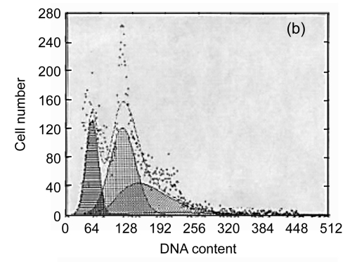
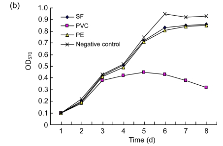
Methods: The effects of regenerated silk fibroin film on cytotoxicity, adhesion, cell cycle, and apoptosis of L929 cells, growth and vascular endothelial growth factor (VEGF) expression of ECV304 cells, and VEGF, angiopoietin-1 (Ang-1), platelet-derived growth factor (PDGF) and fibroblast growth factor 2 (FGF2) expression of WI-38 cells were assessed by 3-(4,5)-dimethylthiahiazo (-z-y1)-3,5-di-phenytetrazoliumromide (MTT) assay, viable cell counting, flow cytometry (FCM), and enzyme-linked immunosorbant assay (ELISA).
Results: We showed that the regenerated silk fibroin film was not cytotoxic to L929 cells and had no adverse influence on their adhesion, cell cycle or apoptosis; it had no adverse influence on the growth and VEGF secretion of ECV304 cells and no effect on the secretion of VEGF, Ang-1, PDGF and FGF2 by WI-38 cells.
Conclusion: The regenerated silk fibroin film should be an excellent biomaterial with good cytocompatibility, providing a framework for reparation after trauma in clinical applications.
Figures


Similar articles
-
[Effect of regenerated silk fibroin film on transcription and expression of vascular endothelial growth factor gene].Sheng Wu Yi Xue Gong Cheng Xue Za Zhi. 2009 Feb;26(1):110-5. Sheng Wu Yi Xue Gong Cheng Xue Za Zhi. 2009. PMID: 19334567 Chinese.
-
Chondrocytes cultured in silk-based biomaterials maintain function and cell morphology.Int J Artif Organs. 2019 Jan;42(1):31-41. doi: 10.1177/0391398818806156. Epub 2018 Oct 30. Int J Artif Organs. 2019. PMID: 30376753
-
Heparinized silk fibroin hydrogels loading FGF1 promote the wound healing in rats with full-thickness skin excision.Biomed Eng Online. 2019 Oct 2;18(1):97. doi: 10.1186/s12938-019-0716-4. Biomed Eng Online. 2019. PMID: 31578149 Free PMC article.
-
Silk Fibroin Biomaterial Shows Safe and Effective Wound Healing in Animal Models and a Randomized Controlled Clinical Trial.Adv Healthc Mater. 2017 May;6(10). doi: 10.1002/adhm.201700121. Epub 2017 Mar 24. Adv Healthc Mater. 2017. PMID: 28337854 Review.
-
[Recent progress on silk fibroin as tissue engineering biomaterials].Zhongguo Xiu Fu Chong Jian Wai Ke Za Zhi. 2008 Feb;22(2):192-5. Zhongguo Xiu Fu Chong Jian Wai Ke Za Zhi. 2008. PMID: 18365617 Review. Chinese.
Cited by
-
Maintaining Inducibility of Dermal Follicle Cells on Silk Fibroin/Sodium Alginate Scaffold for Enhanced Hair Follicle Regeneration.Biology (Basel). 2021 Mar 26;10(4):269. doi: 10.3390/biology10040269. Biology (Basel). 2021. PMID: 33810528 Free PMC article.
-
Silk fibroin and silk-based biomaterial derivatives for ideal wound dressings.Int J Biol Macromol. 2020 Dec 1;164:4613-4627. doi: 10.1016/j.ijbiomac.2020.08.041. Epub 2020 Aug 16. Int J Biol Macromol. 2020. PMID: 32814099 Free PMC article. Review.
-
Beneficial Effects of a Blended Fibroin/Aloe Gel Extract Film on the Biomolecular Mechanism(s) via the MAPK/ERK Pathway Relating to Diabetic Wound Healing.ACS Omega. 2023 Feb 7;8(7):6813-6824. doi: 10.1021/acsomega.2c07507. eCollection 2023 Feb 21. ACS Omega. 2023. PMID: 36844531 Free PMC article.
-
Antimicrobial cryogel dressings towards effective wound healing.Prog Biomater. 2022 Dec;11(4):331-346. doi: 10.1007/s40204-022-00202-w. Epub 2022 Sep 19. Prog Biomater. 2022. PMID: 36123436 Free PMC article. Review.
-
Synthesis and characterization of silk fibroin-MXene composite electrospun fibers for biomedical applications.Front Chem. 2024 Dec 20;12:1471148. doi: 10.3389/fchem.2024.1471148. eCollection 2024. Front Chem. 2024. PMID: 39758157 Free PMC article.
References
-
- Augustin HG, Breier G. Angiogenesis: molecular mechanisms and functional interactions—2nd Kloster Seeon Meeting of the German Priority Research Grant “Angiogenesis”. Thromb Haemos. 2003;89(1):190–197. - PubMed
-
- Buffoni F, Banchelli G, Cambi S. Skin wound healing: some biological parameters in guinea pig. J Pharm Pharmacol. 1993;45:784–790. - PubMed
-
- Freddi G, Romano M, Rosaria M, Tsukada M. Silk fibroin/cellulose blend films: preparation, structure and physical properties. J Appl Polym Sci. 1995;56(12):1537–1545. doi: 10.1002/app.1995.070561203. - DOI
Publication types
MeSH terms
Substances
LinkOut - more resources
Full Text Sources
Research Materials
Miscellaneous
