Review
. 2010 Mar 16;43(3):419-28.
doi: 10.1021/ar900226q.

Bioinspired materials for controlling stem cell fate

Affiliations
Review

Bioinspired materials for controlling stem cell fate

Omar Z Fisher et al. Acc Chem Res. .

Abstract

Although researchers currently have limited ability to mimic the natural stem cell microenvironment, recent work at the interface of stem biology and biomaterials science has demonstrated that control over stem cell behavior with artificial microenvironments is quite advanced. Embryonic and adult stem cells are potentially useful platforms for tissue regeneration, cell-based therapeutics, and disease-in-a-dish models for drug screening. The major challenge in this field is to reliably control stem cell behavior outside the body. Common biological control schemes often ignore physicochemical parameters that materials scientists and engineers commonly manipulate, such as substrate topography and mechanical and rheological properties. However, with appropriate attention to these parameters, researchers have designed novel synthetic microenvironments to control stem cell behavior in rather unnatural ways. In this Account, we review synthetic microenvironments that aim to overcome the limitations of natural niches rather than to mimic them. A biomimetic stem cell control strategy is often limited by an incomplete understanding of the complex signaling pathways that drive stem cell behavior from early embryogenesis to late adulthood. The stem cell extracellular environment presents a miscellany of competing biological signals that keep the cell in a state of unstable equilibrium. Using synthetic polymers, researchers have designed synthetic microenvironments with an uncluttered array of cell signals, both specific and nonspecific, that are motivated by rather than modeled after biology. These have proven useful in maintaining cell potency, studying asymmetric cell division, and controlling cellular differentiation. We discuss recent research that highlights important biomaterials properties for controlling stem cell behavior, as well as advanced processes for selecting those materials, such as combinatorial and high-throughput screening. Much of this work has utilized micro- and nanoscale fabrication tools for controlling material properties and generating diversity in both two and three dimensions. Because of their ease of synthesis and similarity to biological soft matter, hydrogels have become a biomaterial of choice for generating 3D microenvironments. In presenting these efforts within the framework of synthetic biology, we anticipate that future researchers may exploit synthetic polymers to create microenvironments that control stem cell behavior in clinically relevant ways.


Figures

Figure 1
Figure 1
Control Elements of the stem cell niche. The adult stem cell (ASC) can receive both long and short range paracrine and endocrine signals, neural input, and architectural, mechanical and trophic cues from the ECM. Cell-cell communication can occur between the ASC and heterologous niche cells (HNC) or daughter cells (DC). Reprinted by permission from Nature Publishing Group: ref. 4 Copyright 2006.
Figure 2
Figure 2
Biomaterials can be used to modulate the natural stem cell microenvironment. (a) Local delivery of bioactive niche components or inhibitory/stimulatory molecules from a solid (injectable) biomaterial scaffold. (b) Targeting the niche via micro- or nanoparticles that carry and delivery bioactive molecules to manipulate the niche. (c) Local delivery of support cells to augment or manipulate stem cell fates in vivo. Cell delivery could be facilitated using (injectable) biomaterials carriers that likely improve the survival and engraftment of the transplanted cells. (d) Implanted, multicomponent artificial niche that could possibly attract stem cells to populate it (d). Reprinted with permission from Ref. 3 Copyright 2009 Wiley-VCH.
Figure 3
Figure 3
Substrate elasticity directs the differentiation of MSCs. (A) The range of elastic modulus, E for select tissues. (B) MSCs placed on substrates with varied stiffness are initially small and round but overtime change morphology according to substrate elasticity. (i) cell branching per length of primary mouse neurons, MSCs, and blebbistatin-treated MSCs and (ii) spindle morphology of MSCs, blebbistatin-treated MSCs, and mitomycin-C treated MSCs (open squares) compared to C2C12 myoblasts (dashed line). Reprinted with permission from ref. 20 Copyright 2006 Elsevier.
Figure 4
Figure 4
Biomaterial array designed for high throughput analysis of hESC-substrate interactions. (a) monomers used for array synthesis, (b) combinations for the major monomer 1 with monomers A–F, (c) a polymer microarray in triplicate with a close-up of 8 spots, (d) merged fluorescence and laser scattering images showing three representative cell attachments (high, intermediate, low) on the polymer spots. (scale bar = 100mm). (e) Reproduced large-scale stem cell adhesion on polymer film (scale bar = 200mm) Reprinted with permission from ref. 27 Copyright 2009 Wiley-VCH.
Figure 5
Figure 5
Spatially controlled differentiation of mesenchymal stem cells into bony (blue) and fatty tissue (red). Planar cell adhesive micropatterns, such as a square (top left) or an offset annulus (top right) provide controlled regions of high and low cytoskeletal stress, thereby influencing differentiation. Scale bar = 250 μm. (bottom right, left) Multicellular 3D constructs differentiate into a fatty core surrounded by bony tissue, similar to natural long bones. Reprinted with permission from reference 44 Copyright 2008 Wiley-VCH.
Figure 6
Figure 6
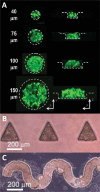
Microfabricated culture wells designed to control the shape of embryoid bodies derived from murine ESCs. (A) Confocal microscopy images of fluorescent labeled EBs within microwells 40–150 in diameter. The technique was also used to create EBs with synthetic shapes, such as triangles (B), and curves (C). Reprinted with permission from ref. 45 Copyright 2007 The Royal Society of Chemistry.
Figure 7
Figure 7
Controlled delivery of retinoic acid (RA) to embryoid bodies. Untreated EBs (A) as well as EBs treated with soluble RA (B), unloaded microspheres (C), or RA-loaded microspheres (D) were formalin-fixed, sectioned and stained with hematoxylin and eosin after 10 days of differentiation. EBs cultured of morphogen loaded microspheres contained a bi-epithelial morphology, with a columnar, pseudo-stratified inner cell layer (black arrows) and an adjacent, flattened outermost cell layer (red arrows). Reprinted with permission from ref. 46 Copyright 2009 Elsevier Inc.
Figure 8
Figure 8
Hydrogel microenvironments with phototunable properties for stem cell encapsulation. The RGDS peptide sequence was coupled to a photodegradable nitrobenzyl ether acrylate to create a photocleavable monomer (A). The monomer was polymerized into a nondegradable gel which upon light exposure releases the tethered peptide (B). Human MSCs were encapsulated in nondegradable PEG gels (b) with or (a) without photoreleasable RGDS. The presentation of RGDS was temporally altered by (c) photocleavage of RGDS from the gel on day 10 in culture. Reprinted with permission from ref. 49 Copyright © 2009 by the American Association for the Advancement of Science.

Similar articles

Cited by

References

    1. Odorico JS, Kaufman DS, Thomson JA. Multilineage differentiation from human embryonic stem cell lines. Stem Cells. 2001;19:193–204. - PubMed
    1. Pittenger MF, Mackay AM, Beck SC, Jaiswal RK, Douglas R, Mosca JD, Moorman MA, Simonetti DW, Craig S, Marshak DR. Multilineage potential of adult human mesenchymal stem cells. Science. 1999;284:143–147. - PubMed
    1. Lutolf MP, Blau HM. Artificial Stem Cell Niches. Advanced Materials. 2009;9999:NA. - PMC - PubMed
    1. Scadden DT. The stem-cell niche as an entity of action. Nature. 2006;441:1075–1079. - PubMed
    1. National Research Council (U.S.) Inspired by biology : from molecules to materials to machines. National Academies Press; Washington, D.C.: 2008. Committee on Biomolecular Materials and Processes. - PubMed

Publication types