. 2010 Feb 3;132(4):1359-70.
doi: 10.1021/ja908562q.

A substructure combination strategy to create potent and selective transthyretin kinetic stabilizers that prevent amyloidogenesis and cytotoxicity

Affiliations

A substructure combination strategy to create potent and selective transthyretin kinetic stabilizers that prevent amyloidogenesis and cytotoxicity

Sungwook Choi et al. J Am Chem Soc. .

Abstract

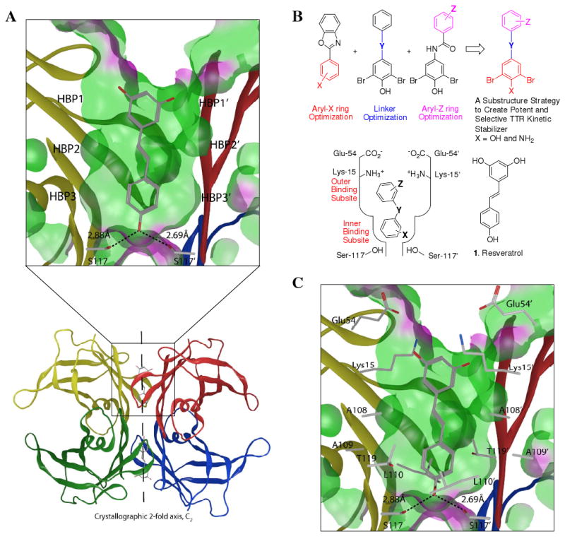
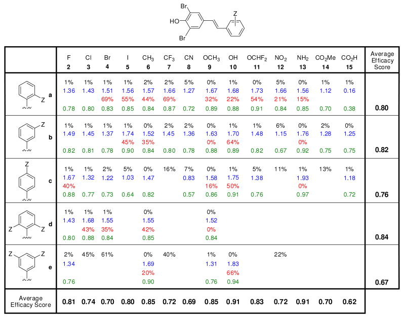
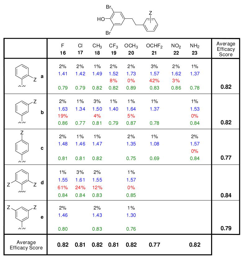
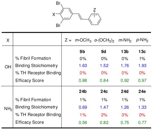
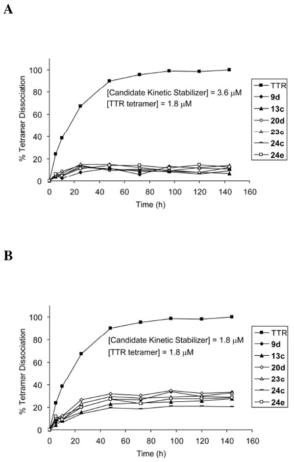
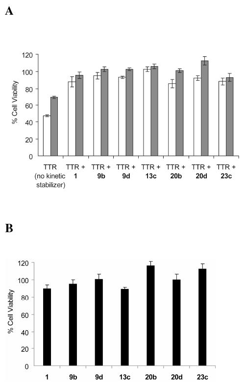
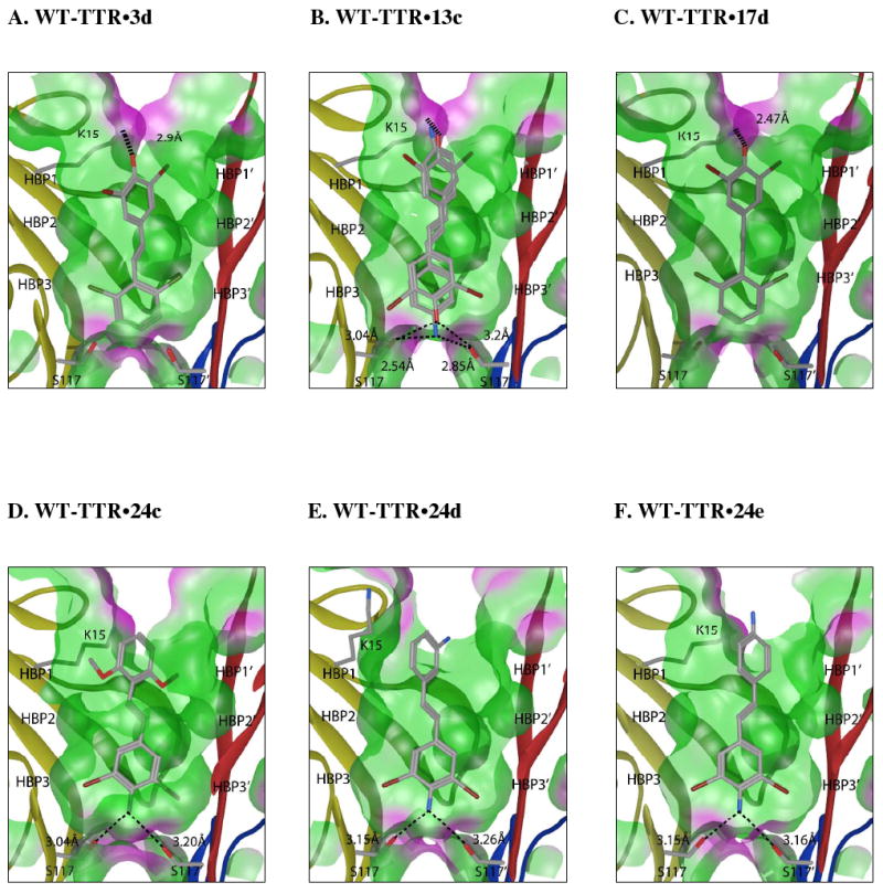
Transthyretin aggregation-associated proteotoxicity appears to cause several human amyloid diseases. Rate-limiting tetramer dissociation and monomer misfolding of transthyretin (TTR) occur before its aggregation into cross-beta-sheet amyloid fibrils. Small molecule binding to and preferential stabilization of the tetrameric state of TTR over the dissociative transition state raises the kinetic barrier for dissociation, imposing kinetic stabilization on TTR and preventing aggregation. This is an effective strategy to halt neurodegeneration associated with polyneuropathy, according to recent placebo-controlled clinical trial results. In three recent papers, we systematically ranked possibilities for the three substructures composing a typical TTR kinetic stabilizer, using fibril inhibition potency and plasma TTR binding selectivity data. Herein, we have successfully employed a substructure combination strategy to use these data to develop potent and selective TTR kinetic stabilizers that rescue cells from the cytotoxic effects of TTR amyloidogenesis. Of the 92 stilbene and dihydrostilbene analogues synthesized, nearly all potently inhibit TTR fibril formation. Seventeen of these exhibit a binding stoichiometry of >1.5 of a maximum of 2 to plasma TTR, while displaying minimal binding to the thyroid hormone receptor (<20%). Six analogues were definitively categorized as kinetic stabilizers by evaluating dissociation time-courses. High-resolution TTR.(kinetic stabilizer)(2) crystal structures (1.31-1.70 A) confirmed the anticipated binding orientation of the 3,5-dibromo-4-hydroxyphenyl substructure and revealed a strong preference of the isosteric 3,5-dibromo-4-aminophenyl substructure to bind to the inner thyroxine binding pocket of TTR.


Figures

Figure 1
Figure 1
Substructure combination strategy to identify potent and selective TTR kinetic stabilizers. (A) Ribbon diagram depiction of the crystal structure (PDB accession code 1DVS) of resveratrol (1) bound to the two thyroxine (T4) binding pockets within the WT-TTR tetramer, with the monomer subunits individually colored. The top portion represents an expanded view of one T4 binding site with 1 bound with a ‘Connelly’ analytical molecular surface applied to residues within 8Å of ligand in the T4 binding pocket (green = hydrophobic, purple = polar). (B) Schematic depiction of the substructure combination strategy to create TTR kinetic stabilizers (top). The aryl-X and aryl-Z rings as well as the linker-Y can be varied to generate candidate TTR kinetic stabilizers. The most highly ranked aryl-X, aryl-Z and linker-Y substructures from previous studies, based on potency and plasma TTR binding stoichiometry, are combined in this substructure combination strategy to create potent and selective TTR kinetic stabilizers that nearly eliminate amyloidogenesis associated cytotoxicity. The inner and outer T4 binding subsites within one schematically represented TTR T4 binding site are labeled in red font. (C) The innermost halogen binding pockets (HBPs) 3 and 3′ (labeled in A) are composed of the methyl and methylene groups of Ser117/117′, Thr119/119′, and Leu110/110′. HBPs 2 and 2′ (labeled in A) are made up by the side chains of Leu110/110′, Ala109/109′, Lys15/15′ and Leu17/17′. The outermost HBPs 1 and 1′ (labeled in A) are lined by the methyl and methylene groups of Lys15/15′, Ala108/108′ and Thr106/106′. Figure generated using the program MOE (2006.08), Chemical Computing Group, Montreal, Canada.
Figure 2
Figure 2
Evaluation of the potency and selectivity of stilbene-based TTR kinetic stabilizers. Percent (%) fibril formation (F.F.) values are in black font representing the extent of in vitro WT-TTR (3.6 μM) fibril formation in the presence of inhibitor (7.2 μM) relative to aggregation in the absence of inhibitor (assigned to be 100%). The TTR tetramer binding stoichiometry of potent aggregation inhibitors (defined as those exhibiting < 10% F.F.) bound to human plasma TTR ex vivo are shown in blue font (10.8 μM kinetic stabilizer concentration, maximum binding stoichiometry is 2 due to the two thyroxine binding sites per TTR tetramer). Extent of competitive binding of potent (based on % F.F. as defined above) and highly selective (defined as those exhibiting a human plasma TTR binding stoichiometry > 1.5) TTR kinetic stabilizers to the thyroid hormone receptor is shown in red font. Individual efficacy scores (as defined by eq. 2) of TTR kinetic stabilizers are shown in green font, whereas average efficacy scores (defined by eq. 1) are shown at the bottom of the columns (reflecting the average value in a column) and at the right side of the rows, reflecting the average value of the compounds in that row.
Figure 3
Figure 3
Evaluation of the potency and selectivity of dihydrostilbene-based WT-TTR kinetic stabilizers. This figure is organized and defined strictly analogous to the descriptions in the legend to Figure 2.
Figure 4
Figure 4
Effect of replacement of a hydroxyl group by an amino group in selected TTR kinetic stabilizers. Percent fibril formation (% F.F.) values (black font), plasma TTR binding stoichiometry (blue font), T3 displacement from thyroid hormone receptor (red font) as well as individual efficacy scores (green font) are shown, precisely as defined in Figure 2, with the exception that average efficacy scores are not shown.
Figure 5
Figure 5
The influence of kinetic stabilizer binding on the rate of TTR tetramer dissociation. WT-TTR (1.8 μM) tetramer dissociation time courses in 6 M urea without (■) and in the presence of putative kinetic stabilizers 9d, 13c, 20d, 23c, 24c, and 24e at a concentration of 3.6 μM (A) and 1.8 μM (B), evaluated by linking the slow tetramer dissociation process to rapid and irreversible monomer denaturation in 6 M urea, as measured by far-UV circular dichroism at 215-218 nm over a time course of 144 h.
Figure 6
Figure 6
(A) Demonstration that kinetic stabilizers of WT-TTR and V30M-TTR can prevent cytotoxicity associated with the process of TTR amyloidogenesis. IMR-32 human neuroblastoma cells were treated with WT-TTR (8 μM, white bar) or V30M-TTR (8 μM, grey bar) exhibiting cytotoxicity that is prevented by preincubating WT-TTR (white bars) or V30M-TTR (grey bars) with the stilbene and dihydrostilbene-based kinetic stabilizers or resveratrol (1) (a kinetic stabilizer previously shown to be effective) included as a positive control (8 μM each). Cell viability was measured after 24 h by the resazurin reduction assay and all the experimental conditions were compared to cells treated with vehicle only (100% cell viability). (B) Cytotoxicity of selected compounds (8 μM) to the human neuroblastoma cell line IMR-32. Columns represent the average values of 2 independently performed experiments (6 experimental replicates). The error bars represent standard error.
Figure 7
Figure 7
Crystal structures of homotetrameric WT-TTR in complex with inhibitors 3d, 13c, 17d, 24c, 24d, and 24e. Ribbon diagram depiction of a close-up view of one of the two identical T4 binding sites (see Figure 1A). A ‘Connelly’ analytical molecular surface was applied to residues within 8Å of ligand in the T4 binding pocket (green = hydrophobic, purple = polar). Polar residues K15 and S117/117′ are shown with bonds depicted where interactions are observed. In the case of stilbene 13c, a mixture of two binding orientations is observed. The 3,5-dibromo-4-hydroxyphenyl ring occupies the thyroxine outer binding subsite ∼90% of the time (see Results section for a more complete description and explanation). Figure generated using the program MOE (2006.08), Chemical Computing Group, Montreal, Canada.

Similar articles

Cited by

References

    1. Balch WE, Morimoto RI, Dillin A, Kelly JW. Science. 2008;319:916–9. - PubMed
    1. Powers ET, Morimoto RI, Dillin A, Kelly JW, Balch WE. Annu Rev Biochem. 2009;78:959–91. - PubMed
    1. Cohen E, Bieschke J, Perciavalle RM, Kelly JW, Dillin A. Science. 2006;313:1604–10. - PubMed
    1. Sekijima Y, Wiseman RL, Matteson J, Hammarstrom P, Miller SR, Sawkar AR, Balch WE, Kelly JW. Cell. 2005;121:73–85. - PubMed
    1. Hammarstrom P, Wiseman RL, Powers ET, Kelly JW. Science. 2003;299:713–6. - PubMed

Publication types