

Evaluation of early cellular influences of bone morphogenetic proteins 12 and 2 on equine superficial digital flexor tenocytes and bone marrow-derived mesenchymal stem cells in vitro
- PMID: 20043789
- PMCID: PMC4246500
- DOI: 10.2460/ajvr.71.1.103
Evaluation of early cellular influences of bone morphogenetic proteins 12 and 2 on equine superficial digital flexor tenocytes and bone marrow-derived mesenchymal stem cells in vitro
Abstract
Objective: To evaluate early cellular influences of bone morphogenetic protein (BMP)12 and BMP2 on equine superficial digital flexor tenocytes (SDFTNs) and equine bone marrow-derived mesenchymal stem cells (BMDMSCs).
Animals: 9 adult clinically normal horses.
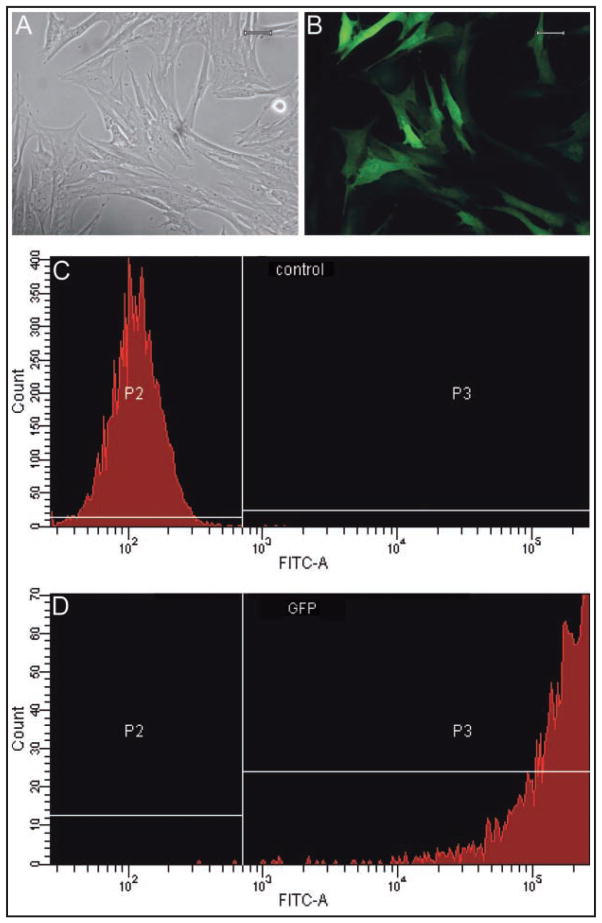
Procedures: BMDMSCs and SDFTNs were cultured in monolayer, either untreated or transduced with adenovirus encoding green fluorescent protein, adenovirus encoding BMP12, or adenovirus encoding BMP2. Cytomorphologic, cytochemical, immunocytochemical, and reverse transcriptase-quantitative PCR (RT-qPCR) analyses were performed on days 3 and 6. Genetic profiling for effects of BMP12 was evaluated by use of an equine gene expression microarray on day 6.
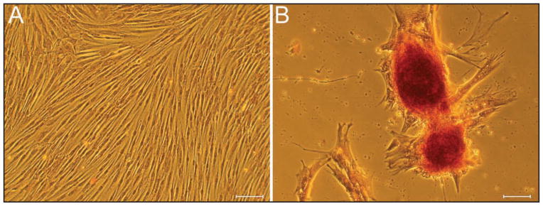
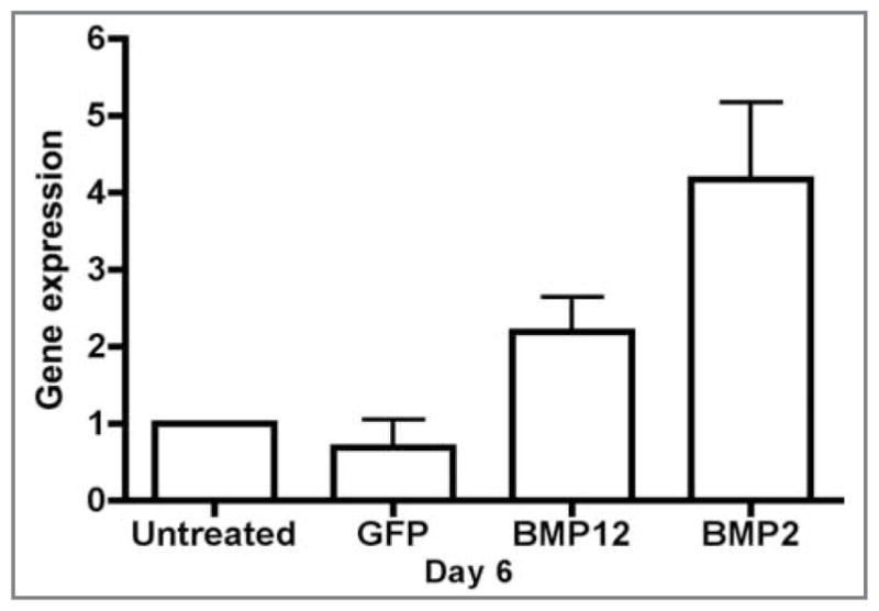
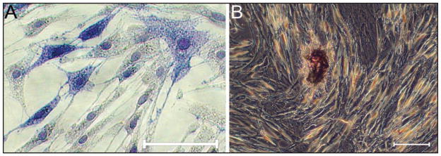
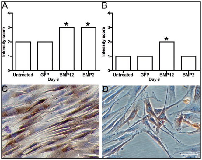
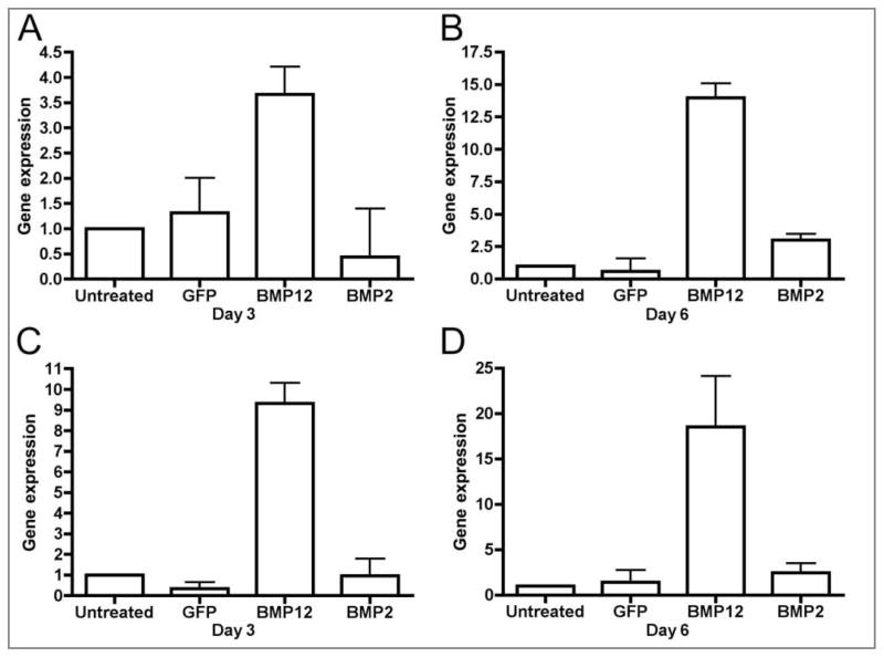
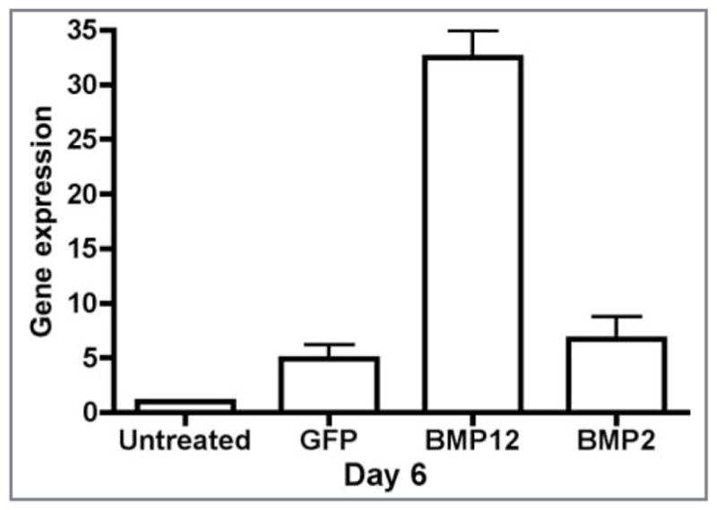
Results: BMDMSCs and SDFTNs had high BMP12 gene expression and remained viable and healthy for at least 6 days. Type l collagen immunocytochemical staining for SDFTNs and tenocyte-like morphology for SDFTNs and BMDMSCs were greatest in BMP12 cells. Cartilage oligomeric matrix protein, as determined via RT-qPCR assay, and chondroitin sulfate, as determined via gene expression microarray analysis, were upregulated relative to control groups in SDFTN-BMP12 cells. The BMDMSCs and SDFTNs became mineralized with BMP2, but not BMP12. Superficial digital flexor tenocytes responded to BMP12 with upregulation of genes relevant to tendon healing and without mineralization as seen with BMP2.
Conclusions and clinical relevance: Targeted equine SDFTNs may respond to BMP12 with improved tenocyte morphology and without mineralization, as seen with BMP2. Bone marrow-derived mesenchymal stem cells may be able to serve as a cell delivery method for BMP12.
Figures

Similar articles
-
Osteoblastic differentiation of human and equine adult bone marrow-derived mesenchymal stem cells when BMP-2 or BMP-7 homodimer genetic modification is compared to BMP-2/7 heterodimer genetic modification in the presence and absence of dexamethasone.J Orthop Res. 2010 Oct;28(10):1330-7. doi: 10.1002/jor.21126. J Orthop Res. 2010. PMID: 20309952 Free PMC article.
-
Growth Factor-Mediated Tenogenic Induction of Multipotent Mesenchymal Stromal Cells Is Altered by the Microenvironment of Tendon Matrix.Cell Transplant. 2018 Oct;27(10):1434-1450. doi: 10.1177/0963689718792203. Epub 2018 Sep 25. Cell Transplant. 2018. PMID: 30251565 Free PMC article.
-
Mesenchymal stem cells differentiate into tenocytes by bone morphogenetic protein (BMP) 12 gene transfer.J Biosci Bioeng. 2005 Oct;100(4):418-22. doi: 10.1263/jbb.100.418. J Biosci Bioeng. 2005. PMID: 16310731
-
Can Extracorporeal Shockwave Promote Osteogenesis of Equine Bone Marrow-Derived Mesenchymal Stem Cells In Vitro?.Stem Cells Dev. 2020 Jan 15;29(2):110-118. doi: 10.1089/scd.2019.0202. Epub 2019 Dec 17. Stem Cells Dev. 2020. PMID: 31744386
-
Gene-mediated osteogenic differentiation of stem cells by bone morphogenetic proteins-2 or -6.J Orthop Res. 2006 Jun;24(6):1279-91. doi: 10.1002/jor.20068. J Orthop Res. 2006. PMID: 16649180
Cited by
-
Culture conditions for equine bone marrow mesenchymal stem cells and expression of key transcription factors during their differentiation into osteoblasts.J Anim Sci Biotechnol. 2013 Oct 29;4(1):40. doi: 10.1186/2049-1891-4-40. J Anim Sci Biotechnol. 2013. PMID: 24169030 Free PMC article.
-
Strategies of tenogenic differentiation of equine stem cells for tendon repair: current status and challenges.Stem Cell Res Ther. 2019 Jun 18;10(1):181. doi: 10.1186/s13287-019-1291-0. Stem Cell Res Ther. 2019. PMID: 31215490 Free PMC article. Review.
-
Interleukin-1β in tendon injury enhances reparative gene and protein expression in mesenchymal stem cells.Front Vet Sci. 2022 Aug 11;9:963759. doi: 10.3389/fvets.2022.963759. eCollection 2022. Front Vet Sci. 2022. PMID: 36032300 Free PMC article.
-
Biological aspects of rotator cuff healing.Muscles Ligaments Tendons J. 2012 Apr 1;1(4):161-8. Print 2011 Oct. Muscles Ligaments Tendons J. 2012. PMID: 23738265 Free PMC article.
-
Modulation of osteoblastic/odontoblastic differentiation of adult mesenchymal stem cells through gene introduction: a brief review.J Korean Assoc Oral Maxillofac Surg. 2013 Apr;39(2):55-62. doi: 10.5125/jkaoms.2013.39.2.55. Epub 2013 Apr 23. J Korean Assoc Oral Maxillofac Surg. 2013. PMID: 24471019 Free PMC article. Review.
References
-
- Williams R, Harkins L, Hammond C, et al. Racehorse injuries, clinical problems and fatalities recorded on British racecourses from flat racing and national hunt racing during 1996, 1997 and 1998. Equine Vet J. 2001;33:478–486. - PubMed
-
- Dyson SJ. Medical management of superficial digital flexor tendonitis: a comparative study of 219 horses (1992–2000) Equine Vet J. 2004;36:415–419. - PubMed
-
- Ross MW. Superficial digital flexor tendonitis. In: Ross MW, Dyson SJ, editors. Diagnosis and management of lameness in the horse. Philadelphia: Saunders; 2003. pp. 628–643.
-
- Dowling BA, Dart AJ, Hodgson DR, et al. Superficial digital flexor tendonitis in the horse. Equine Vet J. 2000;32:369–378. - PubMed
-
- Gillis C. Rehabilitation of tendon and ligament injuries. 43rd Annu Meet Am Assoc Equine Pract. 1997;43:306–309.
Publication types
MeSH terms
Substances
Grants and funding
LinkOut - more resources
Full Text Sources
