. 2009 Dec 31:8:132.
doi: 10.1186/1476-4598-8-132.

Anomalous constitutive Src kinase activity promotes B lymphoma survival and growth

Affiliations

Anomalous constitutive Src kinase activity promotes B lymphoma survival and growth

Jiyuan Ke et al. Mol Cancer. .

Abstract

Background: Previously we have shown that B cell receptor (BCR) expression and B cell receptor signaling pathways are important for the basal growth of B lymphoma cells. In particular we have shown that the activation of Syk, a non-src family protein tyrosine kinase and the mitogen activated protein kinases (MAPK), ERK and JNK that mediate BCR signals are required for the constitutive growth of B lymphoma cells. Since src family protein tyrosine kinases (SFKs) like Lyn are known to be needed for the phosphorylation of BCR co-receptors, Ig-alpha and Ig-beta, we hypothesized that one or more SFKs will be constitutively activated in B lymphoma cells and may be necessary for B lymphoma growth.

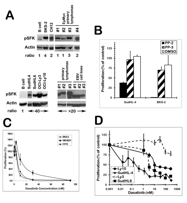
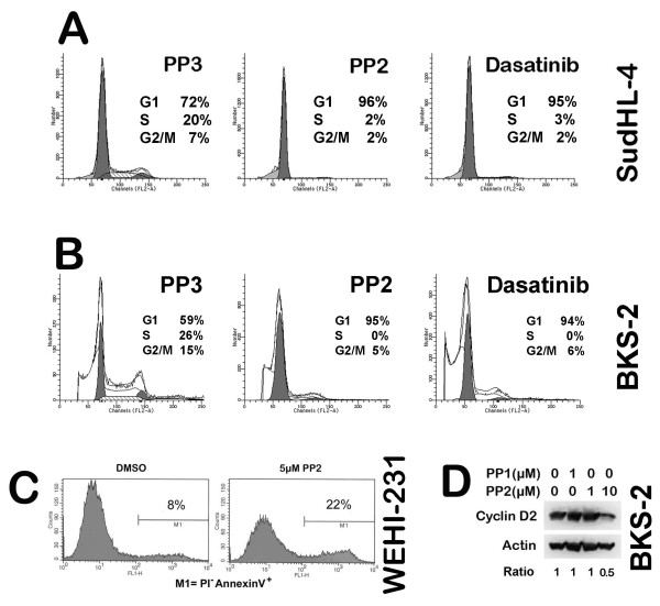
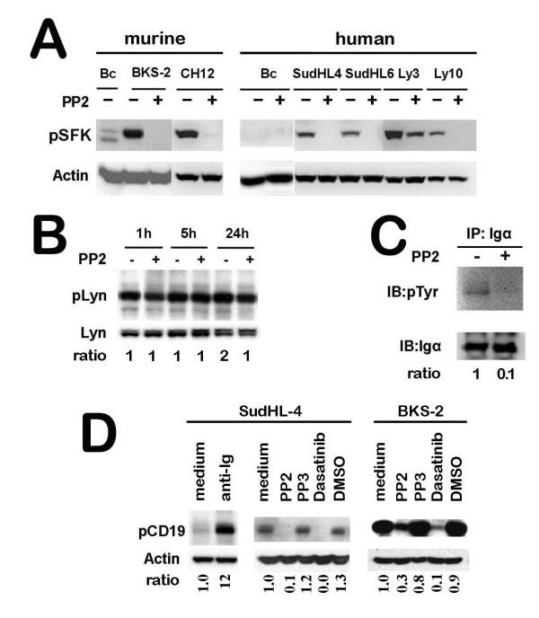
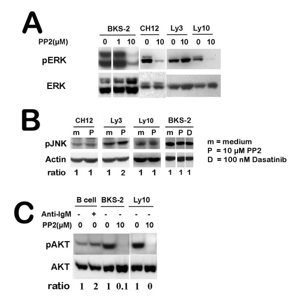
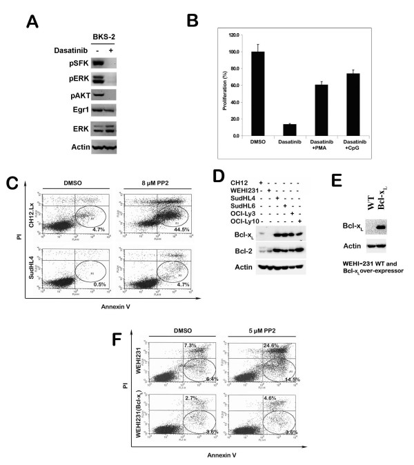
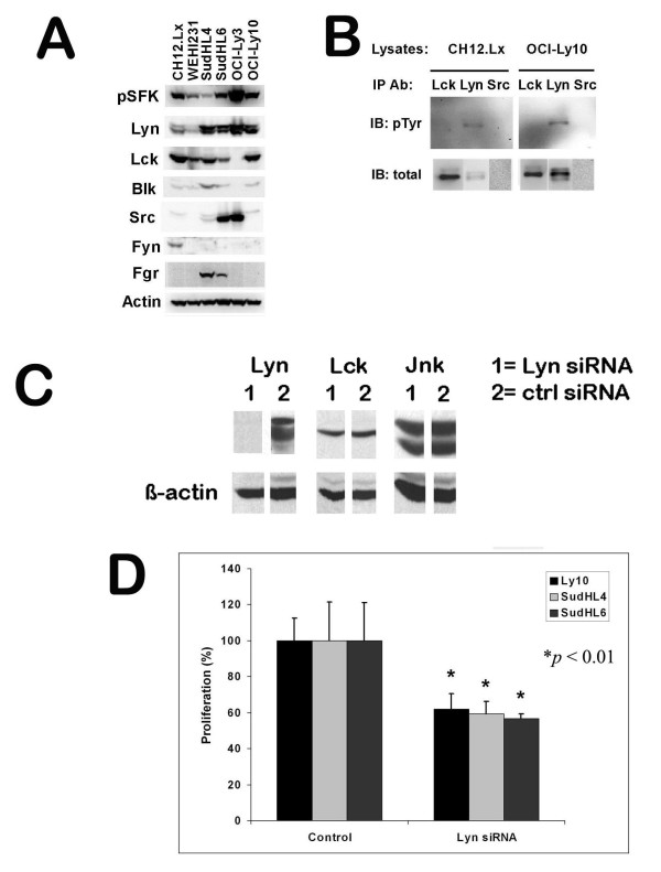
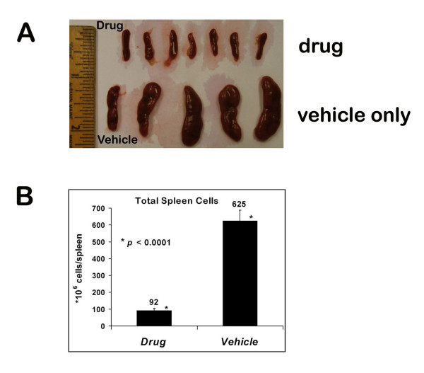
Results: Src kinase activity was found to be constitutively high in many murine and human B lymphoma cell lines and primary lymphoma samples. The specific pharmacological inhibitors of SFKs, PP1 and PP2 inhibited the proliferation of a number of both murine and human B lymphomas in a dose-dependent manner. Importantly, dasatinib (BMS-354825), an oral dual BCR-ABL and SFK specific inhibitor inhibited the growth of B lymphomas in the nanomolar range in vitro and strongly inhibited a mouse lymphoma growth in vivo. Among the SFKs, Lyn is predominantly phosphorylated and Lyn-specific small interfering RNA inhibited the growth of B lymphomas, supporting an important role for Lyn in B lymphoma growth. Suppression of SFK activity blocks BCR mediated signaling pathways. PMA or CpG can partially reverse the growth inhibition induced by SFK inhibition. Although blocking SFK activity inhibited the growth of a number of B lymphomas, some lymphomas such as SudHL-4, SudHL-6, OCI-Ly3 and OCI-Ly10 are more resistant due to an increased expression of the anti-apoptotic proteins Bcl-2 and Bcl-xL.

Conclusions: These studies further support our concept that BCR signaling pathways are important for the continued growth of established B lymphoma cells. Some of the intermediates in this BCR pathway are potential immunotherapeutic targets. In particular, inhibition of SFK activity alone or in synergy with inhibition of the prosurvival Bcl-2 proteins holds promise in developing more effective treatments for B lymphoma patients.


Figures

Figure 1
Figure 1
B lymphomas have high SFK activity as compared to normal B cells. (Panel A) The top row of blots is of mouse and the bottom row is of human B cells. SudHL-4 and 6, Ly3 and 10, and human primary lymphoma #1 are diffuse large B cell lymphomas while the murine primary lymphomas were from EμMyc transgenic mice. Human primary lymphomas #2 & 3 are small cell lymphomas. The cell lysates were first probed for phospho-SFK with p-Src (Y416) antibody, then stripped and re-probed for β-actin as a loading control. The ODs for the p-SFK bands were normalized to the corresponding β-actin bands. The ratios are provided under each lane and are depicted as a fold change over normal cells (i.e. murine lymphomas as a ratio to murine splenic B cells and human lymphomas as a ratio to PB B cells). (B) Cultures of murine (BKS-2) and human (SudHL-4) B lymphomas were treated with the SFK inhibitor PP2 (10 μM) or with the inactive control analog, PP3 (10 μM) for 48 hr and then the proliferation was measured by [3H]-thymidine uptake. Cultures of murine (C) and human (D) B lymphomas were treated with various doses of dasatinib (a dual BCR-ABL and SFK inhibitor) for 48 hr and then the proliferation was measured by [3H]-thymidine uptake. The proliferation assay was performed as described in the "Materials and Methods" section. Data points indicate percent control response ± S.E. of triplicate cultures from a representative experiment. The percent control response is defined as (cpm in the treated group/cpm in the untreated group) × 100. The levels of tritiated thymidine incorporation in the untreated groups were between 34,767 and 84,547 cpm.
Figure 2
Figure 2
PP2 and dasatinib induced G1 cell cycle arrest, accompanied by apoptosis in B lymphoma cells. Cultures of 0.5 × 10⁶/ml human SudHL-4 cells (Panel A) or murine BKS-2 (B) were treated with 10 μM PP2 (SFK inhibitor), PP3 (inactive control analog), or 100 nM dasatinib (dual BCR-ABL and SFK inhibitor) for 48 hr. Then the cells were spun down and stained with PI for cell cycle analysis as described in the Materials and Methods section. (C) 0.5 × 10⁶/ml WEHI-231 cells were treated with 5 μM PP2 or an equivalent amount of DMSO for 48 hrs. Then the cells were spun down and stained with PI and AnnexinV-FITC for apoptosis analysis as described in the Materials and Methods section. (D) BKS-2 cells were treated with or without the indicated amount of PP1 (also a SFK inhibitor) or PP2 for 24 hrs. The cell lysates were probed for cyclin D2, then stripped and re-probed for β-actin. The ODs for cyclin D2 were normalized to the corresponding bands for β-actin and depicted as a fold change over no inhibitor.
Figure 3
Figure 3
PP2 inhibits the phosphorylation of Tyr396 in the activation loop of the kinase domain of Lyn or equivalent position in other SFKs, leading to a disruption of early BCR signaling events. (Panel A) A number of B lymphoma cells were treated with 10 μM PP2 for 1 hr and then cell lysates were prepared for Western blotting. The blot was first probed with p-Src (Y416) antibody, then stripped and re-probed for β-actin. 'Bc' stands for primary B cells. (B) SudHL-4 lymphoma cells were treated with or without 10 μM of PP2 for the indicated time points and cell lysates were prepared for Western blotting. The blot was first probed with p-Lyn (Y507) antibody, then stripped and re-probed for total Lyn. The ODs for p-Lyn were normalized to total Lyn and depicted as a fold change over 1 hr without PP2. (C) SFK inhibition blocks constitutive phosphorylation of Igα in B lymphoma cells. SudHL-4 cells were treated with or without 10 μM PP2 for 1 hr. Igα was immunoprecipitated from equal amount of cell lysates from both groups and analyzed by Western blotting for p-Tyr. The lysates were also probed for Igα as a loading control. The ODs for p-Tyr were normalized to Igα and depicted as a fold change over no PP2 added. (D) SFK inhibition blocks constitutive phosphorylation of CD19 in B lymphoma cells. BKS-2 and SudHL-4 cells were treated as indicated for 4 hr. For a positive control, SudHL-4 cells were treated with 20 μg/ml anti-Ig for 15 min. The cell lysates were analyzed by Western blotting with pCD19 antibody. Then the blots were stripped and re-probed with anti-β-actin antibody. The ODs for pCD19 were normalized to the corresponding β-actin bands and then for each time point, depicted as a fold change over medium only for each panel.
Figure 4
Figure 4
SFK inhibition blocks the phosphorylation of ERK and AKT. (Panel A) Four B lymphoma cells were treated with or without the indicated amount of PP2 for 1 hr. The cell lysates were analyzed by Western blotting for p-ERK. The blot was stripped and re-probed for total ERK. (B) Four B lymphoma cells lines were treated with or without PP2 for 1 hr. The cell lysates were analyzed by Western blotting for p-JNK. The blots were stripped and re-probed for β-actin. The ODs for p-JNK were normalized to the corresponding β-actin bands. For each cell line, the ODs are depicted as a fold change over no PP2. (C) BKS-2 and Ly10 lymphoma cells were treated with or without the indicated amount of PP1 or PP2 for 1 hr. As a positive control, normal splenic B cells were treated with 20 μg/ml anti-IgM for 15 min. The cell lysates were analyzed by Western blotting for p-AKT. The blot was stripped and re-probed for total AKT. The ODs for p-AKT were normalized to the corresponding bands for total AKT. For each cell type, the ODs are depicted as a fold change over no anti-IgM or PP2.
Figure 5
Figure 5
Dasatinib inhibits constitutive BCR signaling and B lymphoma growth. Bcl-2 and Bcl-xL expression confers resistance to PP2 induced apoptosis in B cell lymphomas. (A) Dasatinib inhibits SFK phosphorylation and phosphorylation of its downstream targets ERK and AKT and reduces the level of Egr-1 in BKS-2 lymphoma cells. BKS-2 cells were treated with or without 100 nM dasatinib for 1 hr. The cell lysates were analyzed by Western blotting for p-SFK, p-ERK and p-AKT. The blots were stripped and re-probed for Egr-1, ERK and β-actin. (B) Dasatinib induced growth inhibition can be partially overcome by PMA or CpG. BKS-2 cells were treated with DMSO, 10 nM dasatinib, 10 nM dasatinib+10 ng/ml PMA or 10 nM dasatinib+1 μg/ml CpG for 48 hrs. The proliferation assay was performed as described in the "Materials and Methods" section. Data points indicate percent control response ± S.E. of triplicate cultures from a representative experiment of three independent experiments. The percent control response is defined as (cpm in dasatinib treated group/cpm in DMSO treated group) × 100 for dasatinib; (cpm in dasatinib+PMA treated group/cpm in the PMA treated group) × 100 for dasatinib+PMA; (cpm in dasatinib+CpG treated group/cpm in CpG treated group) × 100 for dasatinib+CpG. The actual counts for DMSO, PMA and CpG treated groups are 16355 ± 1394, 12939 ± 1345, 89504 ± 9159, respectively. (C) SudHL-4 and CH12.Lx cells were treated with or without 8 μM PP2 for two days. Early apoptotic cells were detected by flow cytometry with AnnexinV-APC and PI staining as described in the "Materials and Methods". (D) Bcl-2 and Bcl-xL protein expression in six B lymphoma cells was determined. Cell lysates were analyzed by Western blotting for Bcl-xL and Bcl-2. The blots were stripped and re-probed for β-actin for loading control. (E) The Bcl-xL expression in WEHI-231 and WEHI-231-Bcl-xL cells (WEHI-231 stably transfected with Bcl-xL). Cell lysates were analyzed by Western blotting for Bcl-xL. Then the blot was stripped and re-probed for β-actin for loading control. (F) Ectopic expression of Bcl-xL makes WEHI-231 cells more resistant to PP2 induced apoptosis. WEHI-231 and WEHI-231-Bcl-xL cells were treated with or without 5 μM PP2 for one day. Early apoptotic cells were analyzed by flow cytometry with Annexin-V-FITC and PI staining as described.
Figure 6
Figure 6
Lyn is preferentially expressed, phosphorylated, and plays a critical role for B lymphoma growth. (Panel A) Lyn and Lck are the predominant SFK expressed in a panel of six B lymphoma cell lines. Cell lysates from six representative B lymphoma cell lines were analyzed by Western blotting for p-SFK and different kinds of SFKs. The blots were stripped and re-probed for β-actin for loading control. (B) Lyn is constitutively phosphorylated in CH12.Lx and OCI-Ly10 lymphomas. Lck, Lyn or Src were immunoprecipitated from equal amount of cell lysates from CH12.Lx and OCI-Ly10 and probed for p-Tyr by Western blotting. The blots were then stripped and re-probed for total SFKs. (C) The Lyn siRNA suppressed Lyn expression in SudHL-4 lymphoma cells. SudHL-4 cells were transiently transfected with control or Lyn specific siRNA as described in the Materials and Methods section for 48 hrs. Multiple aliquots of cell lysates were run on SDS-PAGE and transferred onto PVDF membranes. The membranes were cut and separately probed for Lyn, Lck, and Jnk. The blots were stripped and re-probed for the loading control, β-actin. (D) Blocking Lyn expression by siRNA inhibited B lymphoma growth. OCI-Ly10, SudHL-4 and SudHL-6 lymphoma cell lines were treated with control or Lyn specific siRNA as described. Then an equal number of cells with the indicated treatment were used to set up the proliferation assay as described.
Figure 7
Figure 7
Dasatinib inhibits B lymphoma growth in vivo. Dasatinib significantly reduced the splenic B lymphoma burden in CBA/N (Xid) mice. Female CBA/N (Xid) mice were injected intravenously with 7 × 10⁶ BKS-2 B lymphoma cells on day 0. From day 1, mice were injected intraperitoneally either with drug (dasatinib) or vehicle (1 × PBS, 10% DMSO) everyday for 14 days. (A) A comparison of the spleen sizes between drug and vehicle treated lymphoma bearing mice. (B) The average counts of total splenic cells between drug and vehicle treated lymphoma bearing mice.

Similar articles

Cited by

References

    1. Kuppers R. Mechanisms of B-cell lymphoma pathogenesis. Nat Rev Cancer. 2005;5:251–262. doi: 10.1038/nrc1589. - DOI - PubMed
    1. Cattoretti G, Pasqualucci L, Ballon G, Tam W, Nandula SV, Shen Q, Mo T, Murty VV, Dalla-Favera R. Deregulated BCL6 expression recapitulates the pathogenesis of human diffuse large B cell lymphomas in mice. Cancer Cell. 2005;7:445–455. doi: 10.1016/j.ccr.2005.03.037. - DOI - PubMed
    1. Gururajan M, Jennings CD, Bondada S. Cutting edge: constitutive B cell receptor signaling is critical for basal growth of B lymphoma. J Immunol. 2006;176:5715–5719. - PubMed
    1. Kraus M, Alimzhanov MB, Rajewsky N, Rajewsky K. Survival of resting mature B lymphocytes depends on BCR signaling via the Igalpha/beta heterodimer. Cell. 2004;117:787–800. doi: 10.1016/j.cell.2004.05.014. - DOI - PubMed
    1. Alizadeh AA, Eisen MB, Davis RE, Ma C, Lossos IS, Rosenwald A, Boldrick JC, Sabet H, Tran T, Yu X. Distinct types of diffuse large B-cell lymphoma identified by gene expression profiling. Nature. 2000;403:503–511. doi: 10.1038/35000501. - DOI - PubMed

Publication types