. 2009 Dec 31:10:36.
doi: 10.1186/1471-2091-10-36.

Identification of four novel phosphorylation sites in estrogen receptor alpha: impact on receptor-dependent gene expression and phosphorylation by protein kinase CK2

Affiliations

Identification of four novel phosphorylation sites in estrogen receptor alpha: impact on receptor-dependent gene expression and phosphorylation by protein kinase CK2

Christopher C Williams et al. BMC Biochem. .

Abstract

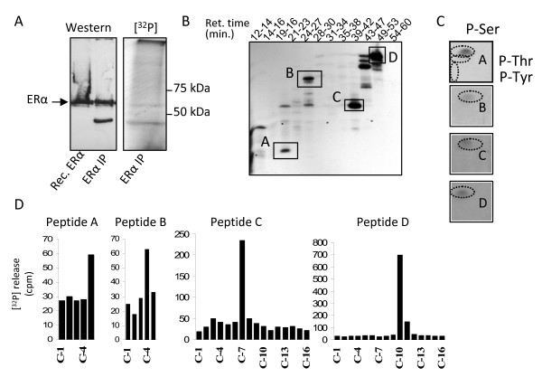
Background: Estrogen receptor alpha (ERalpha) phosphorylation is important for estrogen-dependent transcription of ER-dependent genes, ligand-independent receptor activation and endocrine therapy response in breast cancer. However ERalpha phosphorylation at the previously identified sites does not fully account for these receptor functions. To determine if additional ERalpha phosphorylation sites exist, COS-1 cells expressing human ERalpha were labeled with [32P]H3PO4 in vivo and ERalpha tryptic phosphopeptides were isolated to identify phosphorylation sites.

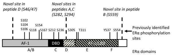
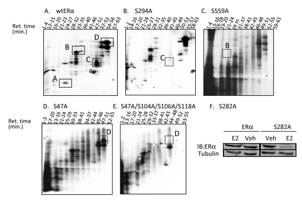
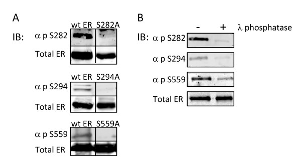
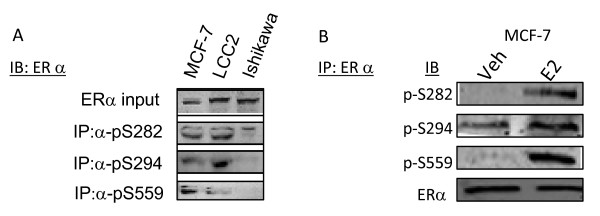
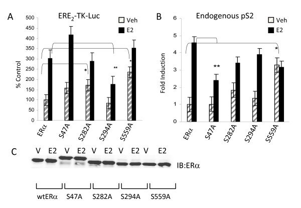
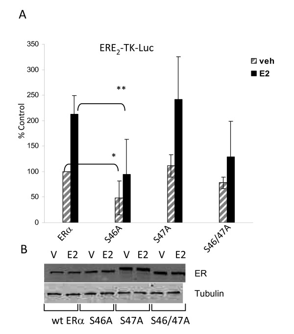
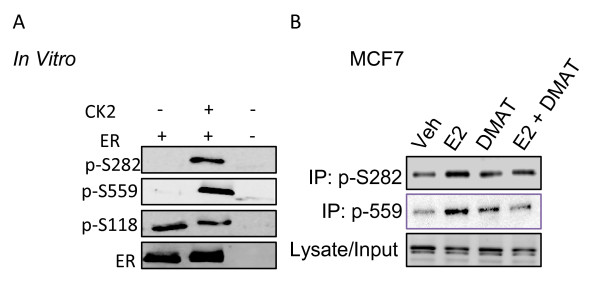
Results: Previously uncharacterized phosphorylation sites at serines 46/47, 282, 294, and 559 were identified by manual Edman degradation and phosphoamino acid analysis and confirmed by mutagenesis and phospho-specific antibodies. Antibodies detected phosphorylation of endogenous ERalpha in MCF-7, MCF-7-LCC2, and Ishikawa cancer cell lines by immunoblot. Mutation of Ser-282 and Ser-559 to alanine (S282A, S559A) resulted in ligand independent activation of ERalpha as determined by both ERE-driven reporter gene assays and endogenous pS2 gene expression in transiently transfected HeLa cells. Mutation of Ser-46/47 or Ser-294 to alanine markedly reduced estradiol dependent reporter activation. Additionally protein kinase CK2 was identified as a kinase that phosphorylated ERalpha at S282 and S559 using motif analysis, in vitro kinase assays, and incubation of cells with CK2 kinase inhibitor.

Conclusion: These novel ERalpha phosphorylation sites represent new means for modulation of ERalpha activity. S559 represents the first phosphorylation site identified in the extreme C-terminus (F domain) of a steroid receptor.


Figures

Figure 1
Figure 1
Estrogen receptor α (ERα) phosphorylation sites. The schematic in Figure 1 depicts both previously identified and novel ERα phosphorylation sites with relative locations within the ERα functional domains. Serines 104, 106, 118, and 167 constitute phosphorylation sites within the ligand-independent activation function-1 (AF-1) domain of ERα. S236 is the first phosphorylation site within the DNA binding domain of ERα. Serine 305, threonine 311 and tyrosine 537 are phosphorylation sites identified within the ligand-dependent activation function-2 (AF-2) domain. Indicated in bold italicized type are newly characterized phosphorylation sites of ERα: S46/47, S282, S294 and S559. S46/47 constitutes an additional site of phosphorylation within the AF-1 domain. Serines 282 and 294 are located in the hinge domain of ERα proximal to the DNA binding domain. Of note, S559 is the first phosphorylation site identified in the extreme C-terminal F domain of ERα and other steroid receptors. S154, S212, S294, S554, and S559 have been recently identified or independently confirmed by mass spectrophotometry (11).
Figure 2
Figure 2
Identification of ERα phosphorylated at serine residues 47, 282, 294 and 559. 6 × 10⁸ COS-1 cells were cultured in phenol red free DMEM supplemented with 10% charcoal stripped FBS. Cells were transfected with wt ERα expression plasmid as noted in materials and methods. 24 hours post-transfection, cells were phosphate-depleted and medium was exchanged with phosphate free DMEM supplemented with 1% dialyzed FBS. 4 mCi [P32] H3PO4 and 10-8 M estradiol were added to each plate and incubated overnight. Cells were subjected to denaturing lysis, ERα purified by immuno-affinity column, eluted, and fractionated by SDS-PAGE. (A) The band corresponding to the 67 kDa ERα was excised and subjected to tryptic digestion. Tryptic ERα peptides were separated by reverse phase HPLC using a C-18 column and 0-45% acetonitrile gradient over 90 minutes. (B) Fractions were collected, pooled according to HPLC retention times and electrophoresed on 40% acrylamide alkaline peptide gels. Gels were autoradiographed, revealing distinct ERα phosphopeptides. 4 novel phosphopeptides (A, B, C, and D) resulting from tryptic digestion of ERα were identified. Each phosphopeptide was then excised, and subjected to modified manual Edman degradation (MED) and phosphoamino acid analysis as described in Materials and Methods. (C) Phosphoamino acid analysis revealed that the phosphopeptide A, B, C and D contained only phosphoserine. Representative phosphoamino acid analyses autoradiograms are presented with identical results detected for phosphopeptides A, B, C, and D. (D) MED detected 32P release for phosphopeptides A-D. Combined data for phosphoamino acid analysis and MED is presented in Table 1.
Figure 3
Figure 3
Mutation of serine residues to alanine eliminates specific phosphorylation of peptides. To confirm the identity of phosphorylated serine residues within peptides A, B, C, and D, serine to alanine mutations were introduced into wt ERα (S47A, S282A, S294A, or S559A). 12 plates of COS-1 cells (4 × 10⁷ /plate) were transfected with 500 ng/plate of wt ERα, S47A, S282A, S294A, or S559A expression plasmids. 18 hours post-transfection, cells were phosphate-depleted, labeled with 4 mCi [32P]H3PO4 and incubated with 10-8 M estradiol overnight. ERα was immunopurified and tryptic peptides were separated by HPLC using a C-18 reversed phase column. Fractions were collected and electrophoresed on a 40% alkaline polyacrylamide gel followed by autoradiography. (A) Peptide map of wt ERα displaying 4 novel phosphopeptides A-D. (B) S294A resulted in loss of peptide C. C) S559A resulted in loss of peptide B. (D) S47A resulted in a modest decrease in peptide D compared to wt ERα. E) S47A/S104A/S106A/S118A resulted in loss of peptide D. (F) Mutation of S282 to alanine reduces ERα protein following 24 h incubation with estradiol. 106 COS-1 monkey embryonic kidney cells which had been cultured in phenol-red free DMEM supplemented with 10% fetal bovine serum were transfected with 2.5 μg of wt ERα or S282A expression plasmid. 24 hours after transfection, cells were incubated with vehicle (veh) or estradiol (10-8M) for an additional 24 hours. Cell lysates were collected and ERα protein levels determined, using α-tubulin as a loading control.
Figure 4
Figure 4
Confirmation of the specificity of ER phospho-antibodies. (A) Serine to alanine mutations at ERα phosphorylation sites inhibit reactivity of phospho-specific antibodies. COS-1 cells cultured in DMEM growth supplemented with 10% FBS were transiently transfected with 500 ng of either wt ERα or serine to alanine substituted ERα expression plasmids (S47A, S282A, S294A, or S559A). 18 hours post-transfection, cells were lysed and subjected to Western immunoblot analysis utilizing custom polyclonal antibodies directed toward the individual phosphorylated ERα residues (S47, S282, S294, or S559) or monoclonal ERα antibody as indicated. α-p-S282, α-p-S294, or α-p-S559 antibodies did not recognize S282A, S294A, or S559A, respectively, indicating phospho-antibody specificity. Mutation of S47 failed to eliminate immunoreactivity of αp-S47. (B) In vitro λ phosphatase treatment of ERα inhibits immunoreactivity of ERα phospho-antibodies. Baculovirus expressed ERα was subjected to dephosphorylation by λ phosphatase for 30 minutes at 30°C and analyzed by Western immunoblot with antibodies against p-S282, p-S294, p-S559, and total ERα. Dephosphorylation inhibited immunoreactivity of, α-p-S282, α-p-S294, and α-p-S559 without impacting immunoreactivity of total ERα antibody.
Figure 5
Figure 5
Phosphorylation of endogenous ERα in ERα (+) cell lines at S282, S294, and S559. (A) 107 MCF-7 and MCF-7-(LCC2) breast cancer and Ishikawa endometrial adenocarcinoma cell lines were cultured in medium supplemented with 10% FBS. Cells were lysed and total ERα was immunoprecipitated with α-p-S282, α-p-S294, or α-p-S559 antibodies for 3 hours. Immunoprecipitates were analyzed by Western blot for total ERα (B) S282 and S559 are phosphorylated following incubation of MCF-7 breast cancer cells with estradiol. MCF-7 breast cancer cells were cultured for 48 hours in phenol red free medium supplemented with 10% charcoal-stripped FBS. Cells were serum starved overnight prior to incubation with vehicle (veh) or 10-8M estradiol (E2). ERα was immunoprecipitated from lysates and Western blot analysis performed with α-pS282, α-pS294, or α-pS559, and with α-ERα. Substantial ligand-induced phosphorylation was detected at S282 and S559, with only modest ligand induced phosphorylation at S294.
Figure 6
Figure 6
Phosphorylation of ERα impacts receptor transcriptional activity. (A) ERα (-) HeLa cells were cotransfected with 100 ng ERE2-TK-luciferase reporter and 200 ng wt ERα (wt), or serine to alanine mutants of ERα for each novel phosphorylation site (S47A, S282A, S294A, and S559A). 24 hours post transfection, cells were incubated with vehicle (veh) or estradiol (10-8 M) overnight. Luciferase assays were performed and transcriptional activity was normalized to protein concentration and/or ERα expression by Western blot analysis. S47A exhibited similar transcriptional activity to wt ERα, whereas S294A resulted in suppressed transcriptional activity vehicle and estradiol. S282A and S559A displayed enhanced ligand independent transcriptional activity as compared to wt ERα. (B) HeLa cervical cancer cells were transfected with 500 ng of wt ERα or ERα phospho-mutant (S47A, S282A, S294A, S559A) expression plasmids. 24 hours post-transfection, cells were incubated with vehicle (veh), 10-8 estradiol (E2), for 3 hours. pS2 expression was measured by real-time RT-PCR, relative to GAPDH. S47A resulted in suppression of estradiol-induced pS2 expression, whereas S559A exhibited ligand-independent activation of ER. S282A and S294A displayed no statistical differences in pS2 mRNA. (C) HeLa cells were transfected with 500 ng of wt ERα or ERα phospho-mutant (S47A, S282A, S294A, and S559A) expression plasmids and incubated for 3 hours with estradiol (1-8M) at 18-24 hours post transfection. Transfection and incubation with estradiol were performed in parallel with those for RT-PCR (panel B). A and B represent composite results for 6 identical experiments. Statistical significance was determined using ANOVA and Fisher's LSD post-hoc analysis, p ≤ 0.05.
Figure 7
Figure 7
Mutation of S46 and S46/47 impacts receptor transcriptional activity. (A) 105 ERα (-) HeLa cells were cotransfected with 100 ng ERE2-TK-luciferase reporter and 200 ng wt ERα (wt), or serine to alanine mutants of ERα for S46 (S46A), S47A (S47A), or both S46 and S47 (S46/47A). 24 hours post transfection, cells were incubated with vehicle (veh) or estradiol (10-8 M) for 18-24 hours. Luciferase assays were performed to determine the relative transcriptional activity of ERα and ERα phospho-mutants. Transcriptional activity was normalized to protein concentration and ERα expression by Western blot analysis. These studies demonstrate that S46A and S46/47A lead to substantial inhibition of ERα mediated gene expression, whereas the activity of S47A remains similar to that of wt ERα. (B) Western blot analysis demonstrating expression of wt ERα, S46A, S47A, and S46/47A. 105 HeLa cells were transfected with 500 ng of wt ERα or ERα phospho-mutant (S46A, S47A, or S46/47A) expression plasmids and incubated for 5 hours with estradiol (1-8M) at 18-24 hours post transfection. S47A shows an electrophoretic upshift not evident with wt ERα, S46A, or S46/47A. Panel A represents the composite of 3 experiments. Statistical significance was determined using ANOVA and Fisher's LSD post-hoc analysis, p ≤ 0.05.
Figure 8
Figure 8
Protein Kinase CK2 phosphorylates of S282 and S559. (A) 400 ng baculovirus expressed ERα was incubated in CK2 kinase buffer supplemented with 10 mM ATP, in the presence or absence of 200 ng recombinant catalytic α subunit of CK2. Reactions were stopped with Laemmli buffer, subjected to Western blot analysis, and probed with α-pS282, α-pS559, α-pS118 or αER. These studies show that the CK2α catalytic subunit specifically phosphorylates ERα at S282 and S559. Western blot for phosphorylation of S118, a site that exhibits strong phosphorylation in baculovirus expressed ERα, is shown for comparison to demonstrate absence of nonspecific phosphorylation by CK2 on other ERα phosphorylation sites. (B) 106 MCF7 breast cancer cells were pretreated with DMAT (4 uM) for 90 minutes, followed by 30 minutes with estradiol (10-8 M) or vehicle. Immunoprecipitation of S282 or S559 was performed using phosphoantibodies and Western blot for total ERα. DMAT inhibited phosphorylation at both sites, indicating that CK2 phosphorylates these sites in vivo.

Similar articles

Cited by

References

    1. Leo C, Chen JD. The SRC family of nuclear receptor coactivators. Gene. 2000;245:1–11. doi: 10.1016/S0378-1119(00)00024-X. - DOI - PubMed
    1. Leo C, Li H, Chen JD. Differential mechanisms of nuclear receptor regulation by receptor-associated coactivator 3. J Biol Chem. 2000;275:5976–5982. doi: 10.1074/jbc.275.8.5976. - DOI - PubMed
    1. Dutertre M, Smith CL. Ligand-Independent Interactions of p160/Steroid Receptor Coactivators and CREB-Binding Protein (CBP) with Estrogen Receptor-{alpha}: Regulation by Phosphorylation Sites in the A/B Region Depends on Other Receptor Domains. Mol Endocrinol. 2003;17:1296–1314. doi: 10.1210/me.2001-0316. - DOI - PubMed
    1. Shah YM, Rowan BG. The Src kinase pathway promotes tamoxifen agonist action in Ishikawa endometrial cells through phosphorylation-dependent stabilization of estrogen receptor (alpha) promoter interaction and elevated steroid receptor coactivator 1 activity. Mol Endocrinol. 2005;19:732–748. doi: 10.1210/me.2004-0298. - DOI - PubMed
    1. Bunone G, Briand PA, Miksicek RJ, Picard D. Activation of the unliganded estrogen receptor by EGF involves the MAP kinase pathway and direct phosphorylation. EMBO J. 1996;15:2174–2183. - PMC - PubMed

Publication types