. 2009 Dec 31:9:303.
doi: 10.1186/1471-2148-9-303.

Conservation of intron and intein insertion sites: implications for life histories of parasitic genetic elements

Affiliations

Conservation of intron and intein insertion sites: implications for life histories of parasitic genetic elements

Kristen S Swithers et al. BMC Evol Biol. .

Abstract

Background: Inteins and introns are genetic elements that are removed from proteins and RNA after translation or transcription, respectively. Previous studies have suggested that these genetic elements are found in conserved parts of the host protein. To our knowledge this type of analysis has not been done for group II introns residing within a gene. Here we provide quantitative statistical support from an analyses of proteins that host inteins, group I introns, group II introns and spliceosomal introns across all three domains of life.

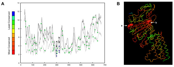
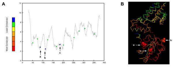
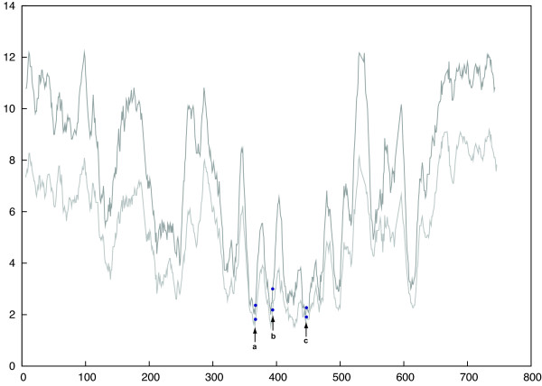
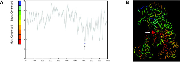
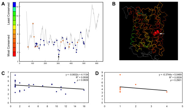
Results: To determine whether or not inteins, group I, group II, and spliceosomal introns are found preferentially in conserved regions of their respective host protein, conservation profiles were generated and intein and intron positions were mapped to the profiles. Fisher's combined probability test was used to determine the significance of the distribution of insertion sites across the conservation profile for each protein. For a subset of studied proteins, the conservation profile and insertion positions were mapped to protein structures to determine if the insertion sites correlate to regions of functional activity. All inteins and most group I introns were found to be preferentially located within conserved regions; in contrast, a bacterial intein-like protein, group II and spliceosomal introns did not show a preference for conserved sites.

Conclusions: These findings demonstrate that inteins and group I introns are found preferentially in conserved regions of their respective host proteins. Homing endonucleases are often located within inteins and group I introns and these may facilitate mobility to conserved regions. Insertion at these conserved positions decreases the chance of elimination, and slows deletion of the elements, since removal of the elements has to be precise as not to disrupt the function of the protein. Furthermore, functional constrains on the targeted site make it more difficult for hosts to evolve immunity to the homing endonuclease. Therefore, these elements will better survive and propagate as molecular parasites in conserved sites. In contrast, spliceosomal introns and group II introns do not show significant preference for conserved sites and appear to have adopted a different strategy to evade loss.


Figures

Figure 1
Figure 1
Positions of inteins and introns along the protein sequence (panel A) and in the structure of vacuolar/archaeal ATPase catalytic subunit (panel B). Panel A shows the conservation profiles of subunit A of the V/A ATPase and the beta subunit of the F-type ATPase. The abscissa shows the amino acid position along the alignment; the ordinate designates the number of different amino acids present in that position averaged over a window of size 11. The dark grey line is the conservation profile of all three domains. The medium gray line is the conservation profile of Eukaryotes and Archaea. The light gray line is the conservation profile of the Archaea. The positions of inteins are indicated as blue dots with arrows. Positions of spliceosomal introns from are indicated as green dots without arrows. Panel B shows the structure of ATPase catalytic subunit A structure from Pyrococcus horikoshii OT3 (PDB ID: 1VDZ[68]) colored according to sequence conservation. The arrows indicate "a" and "b" intein insertion sites. "b" is the archaeal intein insertion site between Lys240 and Thr241 (both amino acids are shown with space-filled model) and "a" is the eukaryotic intein insertion site between Gly260 and Cys261 (space-filled model).
Figure 2
Figure 2
Positions of inteins and spliceosomal introns along the protein sequence (panel A) and in the structure of the replication factor C (panel B). Panel A shows the conservation profile of the RFC protein (see figure 1 for details). The positions of inteins are indicated as blue dots with arrows and positions of spliceosomal introns are green dots without arrows. Panel B shows the structure of the Archaeoglobus fulgidus replication factor C (PDB ID: 2CHV[69]) colored according to sequence conservation. The arrows indicate intein insertion site "a" between Lys51 and THR52, "b" between Ala76 and Ser77, and "c" between Ser138 and Cys139 (all of these six amino acids are shown as space-filled model). All three intein insertion sites are conserved within the host protein.
Figure 3
Figure 3
Positions of inteins along the protein sequence of cell division control protein 21. Dark gray indicates the conservation profile of all domains. Light gray indicates the conservation profile of Eukaryotes only. Inteins are found in three sites "a-c", shown with arrows pointing at blue dots.
Figure 4
Figure 4
Group I intron location along the protein sequence (panel A) and in the structure of the DNA polymerase I (panel B). Panel A shows conservation profile of DNA polymerase I (see figure one for details). Panel B shows the structure of the DNA polymerase I (Klenow Fragment) from Escherichia coli (PDB ID: 1KLN[70]) colored according to site conservation. The arrow indicates intron insertion site, of the intron found in homologous protein in Bacillus subtilis phage SPO1, which is between Asn675 and Leu676 (space-filled model).
Figure 5
Figure 5
Positions of introns along the protein sequence (panel A) and in the structure of the Cytochrome C Oxidase Subunit I (panel B). Panel A shows the conservation profile of the cytochrome C oxidase subunit I (see figure 1 for details). Blue dots indicate group I intron positions and orange dots represent group II intron positions. Panel B displays the structure of the Paracoccus denitrificans cytochrome C oxidase subunit I (PDB ID: 1QLE[71]) colored according to site conservation. The arrow points to the intron insertion site of the intron found in the homologous protein in some vascular plants, protist, fungi, green algae, liverworts, and amoeba. The insertion site is between Gly275 and His276 (space-filled model). Panel C shows the relationship between the number of group I introns found at each position and the site conservation. Panel D shows the relationship between the number of group II introns found at each position and the site conservation.

Similar articles

Cited by

References

    1. Pietrokovski S. Intein spread and extinction in evolution. Trends Genet. 2001;17(8):465–472. doi: 10.1016/S0168-9525(01)02365-4. - DOI - PubMed
    1. Perler FB, Davis EO, Dean GE, Gimble FS, Jack WE, Neff N, Noren CJ, Thorner J, Belfort M. Protein splicing elements: inteins and exteins--a definition of terms and recommended nomenclature. Nucleic Acids Res. 1994;22:1125–1127. doi: 10.1093/nar/22.7.1125. - DOI - PMC - PubMed
    1. Liu XQ. Protein-splicing intein: Genetic mobility, origin, and evolution. Annual review of genetics. 2000;34:61–76. doi: 10.1146/annurev.genet.34.1.61. - DOI - PubMed
    1. Gogarten JP, Senejani AG, Zhaxybayeva O, Olendzenski L, Hilario E. Inteins: structure, function, and evolution. Annu Rev Microbiol. 2002;56:263–287. doi: 10.1146/annurev.micro.56.012302.160741. - DOI - PubMed
    1. Chevalier BS, Stoddard BL. Homing endonucleases: structural and functional insight into the catalysts of intron/intein mobility. Nucleic acids research. 2001;29:3757–3774. doi: 10.1093/nar/29.18.3757. - DOI - PMC - PubMed

Publication types

Substances

LinkOut - more resources