. 2010 Mar 8:1318:133-43.
doi: 10.1016/j.brainres.2009.12.038. Epub 2010 Jan 4.

Lipotoxicity-mediated cell dysfunction and death involve lysosomal membrane permeabilization and cathepsin L activity

Affiliations

Lipotoxicity-mediated cell dysfunction and death involve lysosomal membrane permeabilization and cathepsin L activity

Frankis G Almaguel et al. Brain Res. .

Abstract

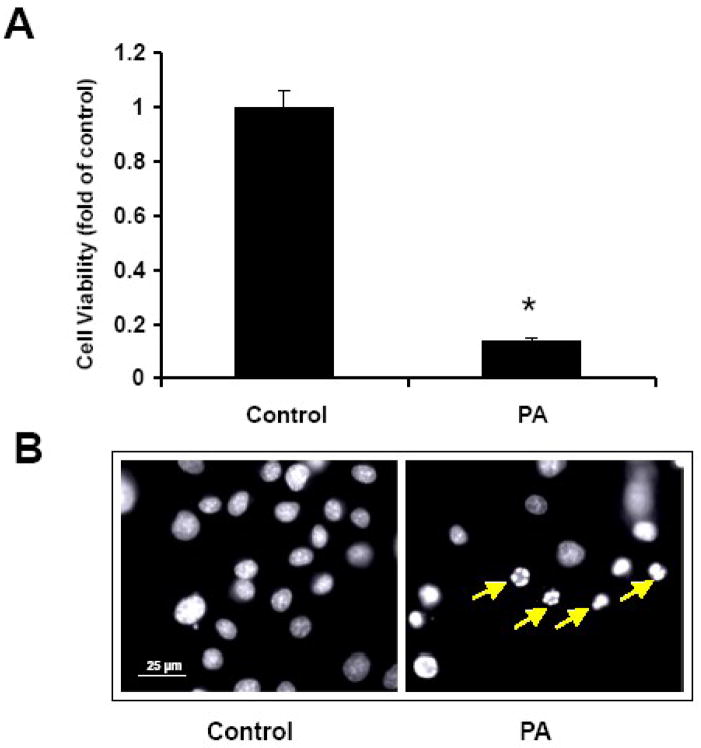
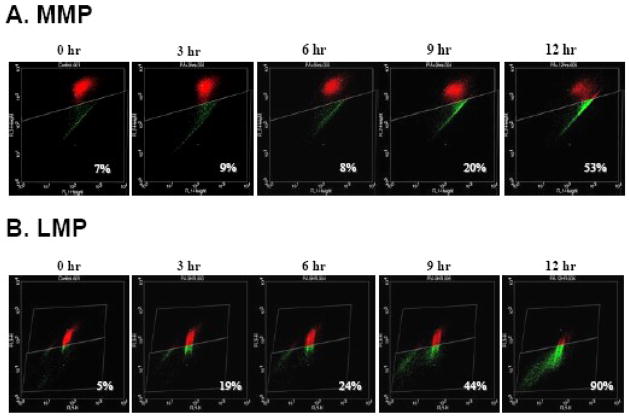
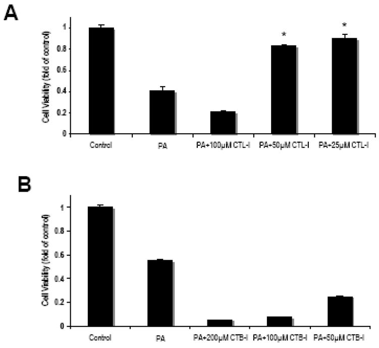
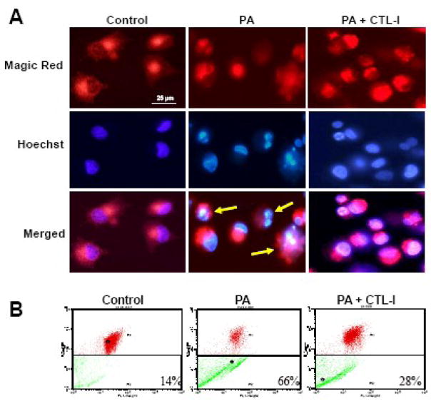
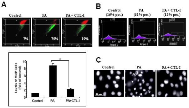
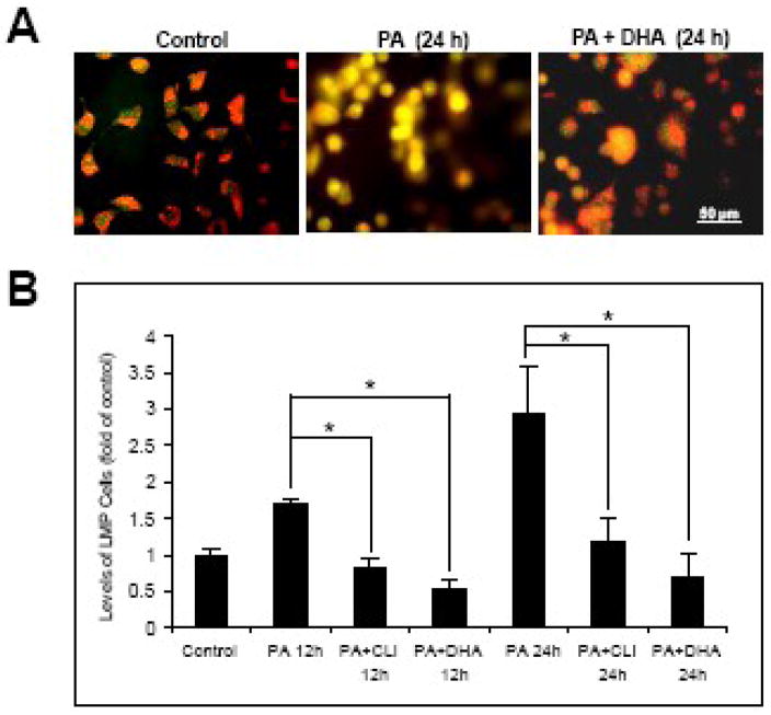
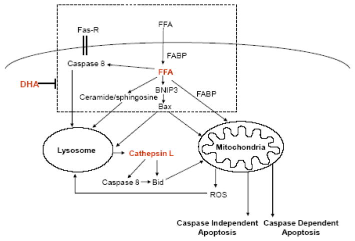
Lipotoxicity, which is triggered when cells are exposed to elevated levels of free fatty acids, involves cell dysfunction and apoptosis and is emerging as an underlying factor contributing to various pathological conditions including disorders of the central nervous system and diabetes. We have shown that palmitic acid (PA)-induced lipotoxicity (PA-LTx) in nerve growth factor-differentiated PC12 (NGFDPC12) cells is linked to an augmented state of cellular oxidative stress (ASCOS) and apoptosis and that these events are inhibited by docosahexanoic acid (DHA). The mechanisms of PA-LTx in nerve cells are not well understood, but our previous findings indicate that it involves ROS generation, mitochondrial membrane permeabilization (MMP), and caspase activation. The present study used nerve growth factor differentiated PC12 cells (NGFDPC12 cells) and found that lysosomal membrane permeabilization (LMP) is an early event during PA-induced lipotoxicity that precedes MMP and apoptosis. Cathepsin L, but not cathepsin B, is an important contributor in this process since its pharmacological inhibition significantly attenuated LMP, MMP, and apoptosis. In addition, co-treatment of NGFDPC12 cells undergoing lipotoxicity with DHA significantly reduced LMP, suggesting that DHA acts by antagonizing upstream signals leading to lysosomal dysfunction. These results suggest that LMP is a key early mediator of lipotoxicity and underscore the value of interventions targeting upstream signals leading to LMP for the treatment of pathological conditions associated with lipotoxicity.


Figures

Figure 1
Figure 1. PA induces lipotoxicity in NGFDPC12 cells
Cells were treated with PA: BSA, 2:1 molar ratio (PA), or BSA alone (Control) for 24 hr. A: Cell viability was determined by WST-1 assay (see Materials and Methods). The data represent mean ± SEM of 3 independent experiments. Significance (*) was determined at p<0.05 compared to control group. B: Nuclear morphology was analyzed with Hoechst staining and examined under fluorescent microscopy. Arrows indicate representative nuclei showing chromatin condensation and fragmentation.
Figure 2
Figure 2. PA induces mitochondrial and lysosomal membrane permeabilization in NGFDPC12 cells
Cells were treated with PA/BSA (2:1) for 0, 3, 6, 9 and 12 hr. A: Mitochondrial membrane permeabilization (MMP) was determined by flow cytometry using JC-1. Cells with MMP showing reduced FL2 reading are presented in green, whereas cells with intact mitochondria are shown in red. The percents of cells with MMP (green) were calculated. B: Lysosomal membrane permeabilization (LMP) was analyzed with flow cytometry using acridine orange (AO). Cells with LMP showing reduced FL2 reading are presented in green, whereas cells with intact lysosomes are shown in red. The percents of cells with LMP (green) were calculated. Representative flow cytometric plots of three independent experiments are shown.
Figure 3
Figure 3. Pharmacological inhibition of cathepsin L attenuates PA-induced cell death in NGFDPC12 cells
A: Cells were treated with PA/BSA (2:1) or BSA alone (Control) for 24 hr in the presence or absence of cathepsin L inhibitor at 25, 50 or 100 μM. Cell viability was determined by WST-1 assay. B: NGFDPC12 cells were treated with PA/BSA (2:1) or BSA alone (CTL) for 24 hr in the presence or absence of cathepsin B inhibitor at 50, 100 or 200 μM. Cell viability was determined by WST-1 assay. The data represent mean ± SEM of 3 independent experiments. Significance (*) was determined at p<0.05 when compared to PA group.
Figure 4
Figure 4
Inhibition of cathepsin L activity attenuates lysosomal membrane permeabilization during PA-induced lipotoxicity in NGFDPC12 cells. Cells were treated with BSA alone (Control), PA/BSA (2:1) or PA/BSA with 25μM cathepsin L inhibitor (PA+CTL-I) for 24 hr. A: Cathepsin L activity in cells was examined with Magic Red and detected by fluorescent microscopy. Cells were counterstained with Hoechst 33342. Merged images are also shown. B: LMP was determined by flow cytometry using acridine orange. The percents of cells with LMP were calculated. Representative flow cytometric plots of three independent experiments are shown.
Figure 5
Figure 5. Cathepsin L inhibitor abolished PA-induced mitochondrial membrane permeabilization and apoptosis
NGFDPC12 cells were treated with BSA alone (Control), PA/BSA (2:1) or PA/BSA with 25μM cathepsin L inhibitor (PA+CTL-I) for 24 hr. A: MMP was determined by flow cytometry using JC-1. The top panel shows flow cytometric plots and the bottom panel shows the quantitative analysis of percent cells with MMP under different treatments. The data represent mean ± SEM of 3 independent experiments. Significance (*) was determined at p<0.05 when compared to PA group. B: Apoptosis was examined by flow cytometry using Annexin V assay, and the percents of Annexin V-positive cells were determined. C: Nuclear morphology after each treatment was visualized with Hoechst 33342 staining. Representative flow cytometric plots and fluorescent micrographs are shown.
Figure 6
Figure 6. DHA protects NGFDPC12 cells against PA-induced lysosomal membrane permeabilization
A: NGFDPC12 cells were treated with BSA alone (Control), PA/BSA (2:1) or PA/BSA+DHA for 24 hr. LMP was determined by acridine orange staining followed by fluorescent microscopy. Representative fluorescent micrographs are shown. B: NGFDPC12 cells were treated with BSA alone (Control), PA/BSA alone, PA/BSA+DHA or PA/BSA with 25μM cathepsin L inhibitor (PA+CLI) for 12 or 24 hr. LMP was analyzed by flow cytometry using Acridine Orange. The percents of cells with LMP were calculated and values for control cells were normalized to one. The data represent mean ± SEM of 3 independent experiments. Significance (*) was determined at p<0.05.
Figure 7
Figure 7. Model depicting the potential lysosomal-mitochondrial crosstalk during saturated free fatty acid-induced apoptosis
FFAs are transported by albumin in the plasma and by fatty acid binding proteins intracellularly. FFA overload leads to: 1) increase of Fas receptor (FAS-R) and activation of caspase 8; 2) enhanced formation of ceramide/sphingosine; and 3) up-regulation of BNIP3 and Bax expression. All these upstream events can contribute to an early lysosomal membrane permeabilization (LMP) and subsequent release of lysosomal enzyme cathepsin L. Released cathepsin L can cleave/activate pro-apoptotic Bid directly or via caspase 8. Translocation of pro-apoptotic proteins to the mitochondrial outer membrane induces mitochondrial membrane permeabilization (MMP), which results in caspase-dependent and independent apoptotic cell death. Reactive oxygen species (ROS) generated by mitochondria can promote LMP and enhance apoptosis. DHA, an n-3 polyunsaturated fatty acid, inhibits FFA-induced apoptosis by blocking upstream signaling cascade and prevents LMP.

Similar articles

Cited by

References

    1. Adibhatla RM, Hatcher JF. Altered lipid metabolism in brain injury and disorders. Subcell Biochem. 2008;49:241–68. - PMC - PubMed
    1. Almaguel FG, Liu JW, Pacheco FJ, Casiano CA, De Leon M. Activation and reversal of lipotoxicity in PC12 and rat cortical cells following exposure to palmitic acid. J Neurosci Res. 2009;87:1207–18. - PMC - PubMed
    1. Antunes F, Cadenas E, Brunk UT. Apoptosis induced by exposure to a low steady-state concentration of H2O2 is a consequence of lysosomal rupture. Biochem J. 2001;356:549–55. - PMC - PubMed
    1. Arab K, Rossary A, Flourié F, Tourneur Y, Steghens JP. Docosahexaenoic acid enhances the antioxidant response of human fibroblasts by upregulating gamma-glutamyl-cysteinyl ligase and glutathione reductase. Br J Nutr. 2006;95:18–26. - PubMed
    1. Bas O, Songur A, Sahin O, Mollaoglu H, Ozen OA, Yaman M, Eser O, Fidan H, Yagmurca M. The protective effect of fish n-3 fatty acids on cerebral ischemia in rat hippocampus. Neurochem Int. 2007;50:548–54. - PubMed

Publication types

MeSH terms