Review
. 2010 Jul;49(3):218-34.
doi: 10.1016/j.plipres.2009.12.003. Epub 2010 Jan 4.

Acyl-coenzyme A binding domain containing 3 (ACBD3; PAP7; GCP60): an emerging signaling molecule

Affiliations
Review

Acyl-coenzyme A binding domain containing 3 (ACBD3; PAP7; GCP60): an emerging signaling molecule

Jinjiang Fan et al. Prog Lipid Res. 2010 Jul.

Abstract

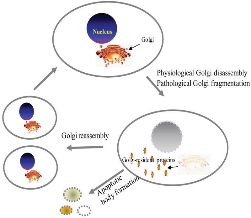
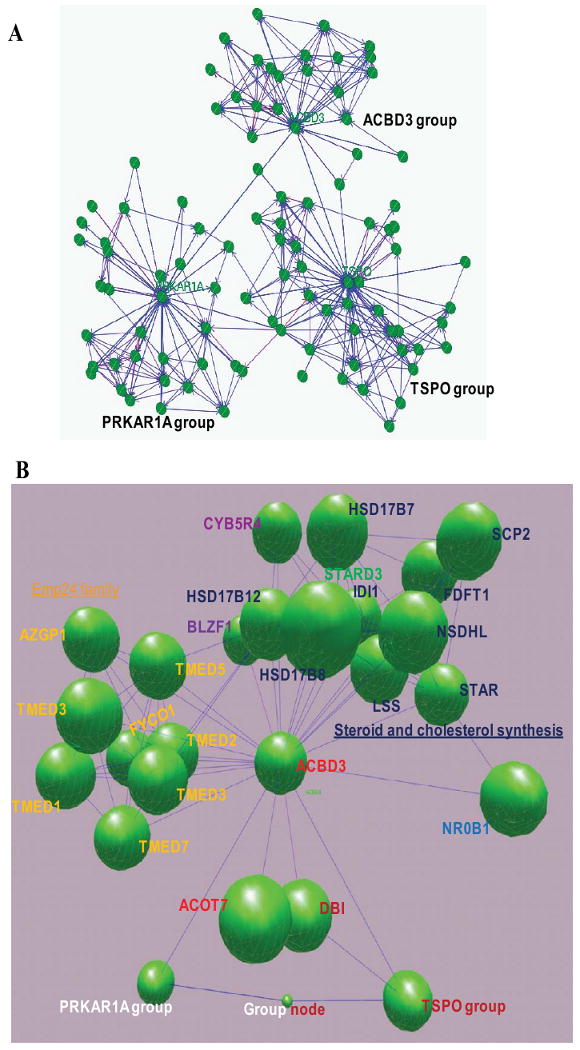
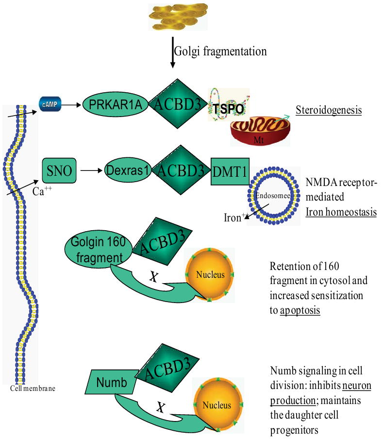
Golgi body-mediated signaling has been linked to its fragmentation and regeneration during the mitotic cycle of the cell. During this process, Golgi-resident proteins are released to the cytosol and interact with other signaling molecules to regulate various cellular processes. Acyl-coenzyme A binding domain containing 3 protein (ACBD3) is a Golgi protein involved in several signaling events. ACBD3 protein was previously known as peripheral-type benzodiazepine receptor and cAMP-dependent protein kinase associated protein 7 (PAP7), Golgi complex-associated protein of 60kDa (GCP60), Golgi complex-associated protein 1 (GOCAP1), and Golgi phosphoprotein 1 (GOLPH1). In this review, we present the gene ontology of ACBD3, its relations to other Acyl-coenzyme A binding domain containing (ACBD) proteins, and its biological function in steroidogenesis, apoptosis, neurogenesis, and embryogenesis. We also discuss the role of ACBD3 in asymmetric cell division and cancer. New findings about ACBD3 may help understand this newly characterized signaling molecule and stimulate further research into its role in molecular endocrinology, neurology, and stem cell biology.


Figures

Fig. 1
Fig. 1
Schematic illustration of Golgi apparatus fragmentation and reassembly during cell mitosis and apoptosis. Many reasons can lead to Golgi fragmentation, but it can be generally classified as physiological Golgi disassembly (Mitosis) or pathological Golgi fragmentation due to apoptosis, presence of misfolded proteins, neurodegenerative diseases, Chlamydia infection, etc.
Fig. 2
Fig. 2
Schematic domain architecture and sequence alignment of ACBD3 and other ACBP domain-containing proteins in humans. A. Organization of ACBP domain-containing proteins. ACBD3 contains an ACBP domain (shown in green) at the N-terminus, a GOLD (for for Golgi dynamics) domain at the C-terminus, and a putative nuclear localization signal (NLS) located between the ACBP- and GOLD- domains. ACBD2 has one enoyl-CoA hydratase/isomerase (ECH) domain closer to the C-terminus. ACBD6 possesses two Ankyrin repeats (Ank) at the C-terminus. B. The positions of the four alpha-helices are indicated (H1-4). Red arrowheads indicate the important binding sites for acyl-CoA type ligands [62]. The conserved sites are in red with black background. C. A neighbor-joining (NJ) tree calculated from the amino acid sequences of human and mouse ACBP domain-containing proteins. The numbers at each node indicate the percentage of bootstrap values from 1000 resamplings. Bar indicates that the estimated genetic distance is proportional to the horizontal length of each branch.
Fig. 2
Fig. 2
Schematic domain architecture and sequence alignment of ACBD3 and other ACBP domain-containing proteins in humans. A. Organization of ACBP domain-containing proteins. ACBD3 contains an ACBP domain (shown in green) at the N-terminus, a GOLD (for for Golgi dynamics) domain at the C-terminus, and a putative nuclear localization signal (NLS) located between the ACBP- and GOLD- domains. ACBD2 has one enoyl-CoA hydratase/isomerase (ECH) domain closer to the C-terminus. ACBD6 possesses two Ankyrin repeats (Ank) at the C-terminus. B. The positions of the four alpha-helices are indicated (H1-4). Red arrowheads indicate the important binding sites for acyl-CoA type ligands [62]. The conserved sites are in red with black background. C. A neighbor-joining (NJ) tree calculated from the amino acid sequences of human and mouse ACBP domain-containing proteins. The numbers at each node indicate the percentage of bootstrap values from 1000 resamplings. Bar indicates that the estimated genetic distance is proportional to the horizontal length of each branch.
Fig. 3
Fig. 3
Comparison of the 3D structures of ACBP domains from each ACBP domain-containing protein. Left panel: Ribbon presentation of 3D models of each ACBP domain. The five conserved amino acids comprising the acyl-CoA binding sites are presented in a ball-and stick model with CPK colors. Right panel: Electrostatic potentials are mapped on the protein surface colored from red (negative) to blue (positive) and yellow (hydrophobic) using eF-Surf service (http://ef-site.hgc.jp/eF-surf) and jV Version 3.6 [109]. The positive binding cavity is shown. Crystal structures are from human ACBD1 (2FJ9), ACBD2 (2CQU) and ACBD6 (2COP). Homology models are deduced via the Swiss-model services using NMR- and/or crystal structures of human ACBD1 and ACBD6 (2FJ9 and 2COP) as templates.
Fig. 4
Fig. 4
Electrostatic potential of the ACBP domains from human ACBD3 (left) and ACBD1 (right), superimposed with a stick representation of the palmitoyl-coenzyme A ligand (in yellow) and backbone of bovine ACBP as a template (PDB: 1ACA) [62]. The electrostatic molecular surfaces are colored by electrostatic potential: red, negatively charged regions, blue, positively charged areas, and white, hydrophobic surface. The four/five residues, which are necessary for ligand binding, are indicated in the figure [63].
Fig. 5
Fig. 5
Multiple alignment of ACBD3 proteins from vertebrates. Sequences were aligned using the ClusterW algorithm. Sequences are designated by species and were obtained from Genbank: human (NP_073572), mouse (NP_573488), rat (AAH83877), pan (gorrila; XP_001140648), bovine (XP_001251579), macaca (monkey; XP_001091471), Monodelphis (opossum; XP_001368267), Gallus (chicken; NP_001026214), frog (AAI35954), and danio (zebrafish; CAI21107). Shaded areas represent conserved amino acids. The numbers indicate amino acid positions. Conserved amino acids are indicated by upper case letters, while less conserved amino acids are represented by lower case letters at the foot of the alignment. The two main domains, ACBP and GOLD, are indicated with underlines. Two putative nuclear localization signals are boxed in red. The five putative acyl-CoA ligand-binding residues are marked with arrowheads. The green boxes indicate the N0 isoform of D-AKAP1 targeting domain 1–30, human PAP7 peptide residue 68–97, the dual type I and type II domain (317–338) of D-AKAP1, and hPAP7 238–259. Green arrows indicate conserved sites, as shown previously. The purple arrow indicates the location of cys463 in human ACBD3 that interacts with golgin-160-(140–311) in a redox dependent reaction.
Fig. 6
Fig. 6
Neighbor-joining (NJ) trees of the ACBP domain, the GOLD domain from ACBD3 proteins, and other domain-containing proteins. A. ACBP domain-containing proteins. ACBP domain-containing proteins (other than ACBD3) are in a compressed branch with a strong bootstrap support (99% from 1000 resamplings). B. GOLD domain-containing proteins. The EMP24 family (integral membrane components of endoplasmic reticulum-derived COPII-coated vesicles) is clearly in a separated compressed clade in this NJ tree, suggesting that the two main branches are present in the GOLD/EMP24 family. The number at each node represents the percentage of NJ bootstrap values from 1000 replicates. The estimated genetic distance is proportional to the horizontal length of each branch. The GenBank accession numbers and other abbreviations used here are listed in Table 2.
Fig. 7
Fig. 7
Redistribution of ACBD3 after MA-10 cells were treated with hCG (2), H-89 (3), and hCG/H-89 (4). The MA-10 cells transfected with plasmid ACBD3-eGFP without any treatment was used as control (1). The vesicle docking protein p115 was used as a marker of Golgi body, while DAPI staining marked the nucleus.
Fig. 8
Fig. 8
ACBD3, a central node in steroidogenesis. A. Gene networks were constructed through the IntNetDB server (http://hanlab.genetics.ac.cn/IntNetDB.htm) and visualized by BioLayout (http://www.biolayout.org) using human ACBD3 (Gene ID: 64746), TSPO (Gene ID: 706), and PRKAR1A (Gene ID: 5573) genes as probes, respectively. B. A 3D graph of the ACBD3 gene network, with collapsed TSPO- and PRKAR1A- groups. Steroid and cholesterol synthesis-related genes (in green) in the ACBD3 gene network: STAR, steroidogenic acute regulatory protein; SCP2, sterol carrier protein 2; FDFT1, farnesyl-diphosphate farnesyltransferase 1 (squalene synthase); NSDHL, NAD(P) dependent steroid dehydrogenase-like; LSS, lanosterol synthase (2,3-oxidosqualene-lanosterol cyclase); IDI1, isopentenyl-diphosphate delta isomerase 1; STARD3, StAR-related lipid transfer (START) domain containing 3; HSD17B7, hydroxysteroid (17-beta) dehydrogenase 7; HSD17B12, hydroxysteroid (17-beta) dehydrogenase 12; HSD17B8, hydroxysteroid (17-beta) dehydrogenase 8; CYB5R4, cytochrome b5 reductase 4 (N-terminal cytochrome b5 and cytochrome b5 oxidoreductase). Hormone nuclear receptor (in dark blue): NR0B1, nuclear receptor subfamily 0, group B, member 1. Acyl coenzyme-related: DBI, diazepam binding inhibitor (GABA receptor modulator, acyl-Coenzyme A binding protein); BACH (ACOT7), acyl-CoA thioesterase 7 (brain acyl-CoA hydrolase). Golgi proteins: BLZF1, basic leucine zipper nuclear factor 1 (JEM-1). Lipid degradation: AZGP1, alpha-2-glycoprotein 1, zinc-binding. Emp24 family (in yellow): TMED1, transmembrane emp24 protein transport domain containing 1; TMED9, transmembrane emp24 protein transport domain containing 9; TMED5, transmembrane emp24 protein transport domain containing 5; TMED3, transmembrane emp24 protein transport domain containing 3; RNP24, transmembrane emp24 domain trafficking protein 2 (transmembrane emp24 domain trafficking protein 2); TMED7, transmembrane emp24 protein transport domain containing 7; FYCO1, FYVE and coiled-coil domain containing 1.
Fig. 9
Fig. 9
ACBD3-mediated signaling pathway and cellular regulation. Following release after Golgi fragmentation, ACBD3 interacts with several other signaling molecules. ACBD3 then regulates diverse cellular processes, including steroidogenesis, iron homeostasis, apoptosis, and neurogenesis. ACBD3 serves as an intermediate, and may be involved in the signaling protein complex related to steroidogenesis and NMDA receptor-mediated iron homeostasis. ACBD3 holds a Golgi 160 fragment and numb protein within the cytoplasm. ACBD3 then regulates cell apoptosis and neurogenesis. Mt, mitochondria; TSPO, translocator protein, 18 kDa; PRKAT1A, protein kinase, cAMP-dependent, regulatory, type I, alpha (tissue specific extinguisher 1); cAMP, cyclic adenosine monophosphate; DMT1, divalent metal transporter 1; Dexras1, a 30 kDa brain-enriched member of the Ras family of small monomeric G proteins; SNO, S-nitrosothiol; Golgin-160 fragment, caspase cleavaged fragment of the golgin family of Golgi-localized proteins; Numb, numb homolog (Drosophila).

Similar articles

Cited by

References

    1. Barr FA. Golgi inheritance: shaken but not stirred. J Cell Biol. 2004;164:955–958. - PMC - PubMed
    1. Misumi Y, Sohda M, Ikehara Y. GCP60, a novel Golgi phosphoprotein, interact with giantin and involved in the maintenance of the Golgi structure. Cell Struct Funct. 2000;25:410.
    1. Sohda M, Misumi Y, Yamamoto A, Yano A, Nakamura N, Ikehara Y. Identification and characterization of a novel Golgi protein, GCP60, that interacts with the integral membrane protein giantin. J Biol Chem. 2001;276:45298–45306. - PubMed
    1. Barr FA, Short B. Golgins in the structure and dynamics of the Golgi apparatus. Curr Opin Cell Biol. 2003;15:405–413. - PubMed
    1. Li H, Degenhardt B, Tobin D, Yao ZX, Tasken K, Papadopoulos V. Identification, localization, and function in steroidogenesis of PAP7: a peripheral-type benzodiazepine receptor- and PKA (RIalpha)-associated protein. Mol Endocrinol. 2001;15:2211–2228. - PubMed

Publication types

MeSH terms

Substances