

NG2 cells: Properties, progeny and origin
- PMID: 20043946
- PMCID: PMC2862831
- DOI: 10.1016/j.brainresrev.2009.12.006
NG2 cells: Properties, progeny and origin
Abstract
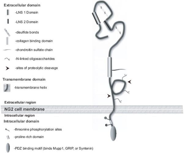
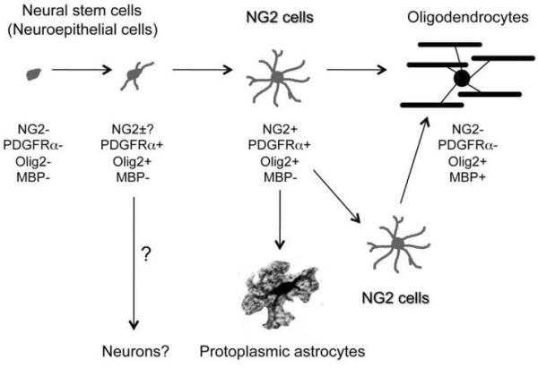
The NG2 proteoglycan is a type 1-transmembrane protein expressed by a range of cell types within and outside the mammalian nervous system. NG2-expressing (NG2) cells are found in grey and white matter tracts of the developing and adult CNS and have previously been assumed to represent oligodendrocyte precursor cells: new work using transgenic mice has shown that NG2 cells generate oligodendrocytes, protoplasmic astrocytes and in some instances neurons in vivo. NG2 cells express GABAA receptors and the AMPA subtype of glutamate receptors. They make intimate contact to neurons prior to myelinating axons and also form electron-dense synaptic specialisations with axons in the cerebellum, cortex and hippocampus and with non-myelinated axons in the corpus callosum. These synaptic NG2 cells respond to neuronal release of glutamate and GABA. This neuron-glia interaction may thus regulate the differentiation and proliferation of NG2 cells. The C-terminal PDZ-binding motif of the NG2 protein binds several PDZ proteins including Mupp1, Syntenin and the Glutamate Receptor Interacting Protein (GRIP). Since GRIP can bind subunits of the AMPA receptors expressed by NG2 cells, the interaction between GRIP and NG2 may orientate the glial AMPA receptors towards sites of neuronal glutamate release. The origin, heterogeneity and function of NG2 cells as modulators of the neuronal network are important incompletely resolved questions.
Copyright 2009 Elsevier B.V. All rights reserved.
Figures

Similar articles
-
The proteoglycan NG2 is complexed with alpha-amino-3-hydroxy-5-methyl-4-isoxazolepropionic acid (AMPA) receptors by the PDZ glutamate receptor interaction protein (GRIP) in glial progenitor cells. Implications for glial-neuronal signaling.J Biol Chem. 2003 Feb 7;278(6):3590-8. doi: 10.1074/jbc.M210010200. Epub 2002 Nov 27. J Biol Chem. 2003. PMID: 12458226
-
Synapses between NG2 glia and neurons.J Anat. 2011 Jul;219(1):2-7. doi: 10.1111/j.1469-7580.2011.01359.x. Epub 2011 Mar 13. J Anat. 2011. PMID: 21395579 Free PMC article. Review.
-
NG2-expressing cells in the nervous system: role of the proteoglycan in migration and glial-neuron interaction.J Anat. 2005 Dec;207(6):735-44. doi: 10.1111/j.1469-7580.2005.00461.x. J Anat. 2005. PMID: 16367801 Free PMC article. Review.
-
Neuron-glia synapses in the brain.Brain Res Rev. 2010 May;63(1-2):130-7. doi: 10.1016/j.brainresrev.2009.12.003. Epub 2009 Dec 16. Brain Res Rev. 2010. PMID: 20018210 Free PMC article. Review.
-
The fate of synaptic input to NG2 glial cells: neurons specifically downregulate transmitter release onto differentiating oligodendroglial cells.J Neurosci. 2010 Jun 16;30(24):8320-31. doi: 10.1523/JNEUROSCI.0854-10.2010. J Neurosci. 2010. PMID: 20554883 Free PMC article.
Cited by
-
Delayed cell cycle pathway modulation facilitates recovery after spinal cord injury.Cell Cycle. 2012 May 1;11(9):1782-95. doi: 10.4161/cc.20153. Epub 2012 May 1. Cell Cycle. 2012. PMID: 22510563 Free PMC article.
-
Chondroitin Sulfate Proteoglycan 4 as a Marker for Aggressive Squamous Cell Carcinoma.Cancers (Basel). 2022 Nov 13;14(22):5564. doi: 10.3390/cancers14225564. Cancers (Basel). 2022. PMID: 36428658 Free PMC article. Review.
-
Chondroitin Sulphate Proteoglycan Axonal Coats in the Human Mediodorsal Thalamic Nucleus.Front Integr Neurosci. 2022 Jul 6;16:934764. doi: 10.3389/fnint.2022.934764. eCollection 2022. Front Integr Neurosci. 2022. PMID: 35875507 Free PMC article.
-
Fibroblast growth factor signaling is required for the generation of oligodendrocyte progenitors from the embryonic forebrain.J Neurosci. 2011 Mar 30;31(13):5055-66. doi: 10.1523/JNEUROSCI.4800-10.2011. J Neurosci. 2011. PMID: 21451043 Free PMC article.
-
Evolution of glial cells: a non-bilaterian perspective.Neural Dev. 2024 Jun 21;19(1):10. doi: 10.1186/s13064-024-00184-4. Neural Dev. 2024. PMID: 38907299 Free PMC article. Review.
References
-
- Anderson SA, Marin O, Horn C, Jennings K, Rubenstein JL. Distinct cortical migrations from the medial and lateral ganglionic eminences. Development. 2001;128:353–63. - PubMed
-
- Bergles DE, Roberts JD, Somogyi P, Jahr CE. Glutamatergic synapses on oligodendrocyte precursor cells in the hippocampus. Nature. 2000;405:187–91. - PubMed
Publication types
MeSH terms
Substances
Grants and funding
LinkOut - more resources
Full Text Sources
Other Literature Sources
Medical
