. 2010 Apr;22(4):684-96.
doi: 10.1016/j.cellsig.2009.12.003. Epub 2010 Jan 5.

Agonist-dependent mu-opioid receptor signaling can lead to heterologous desensitization

Affiliations

Agonist-dependent mu-opioid receptor signaling can lead to heterologous desensitization

Ji Chu et al. Cell Signal. 2010 Apr.

Abstract

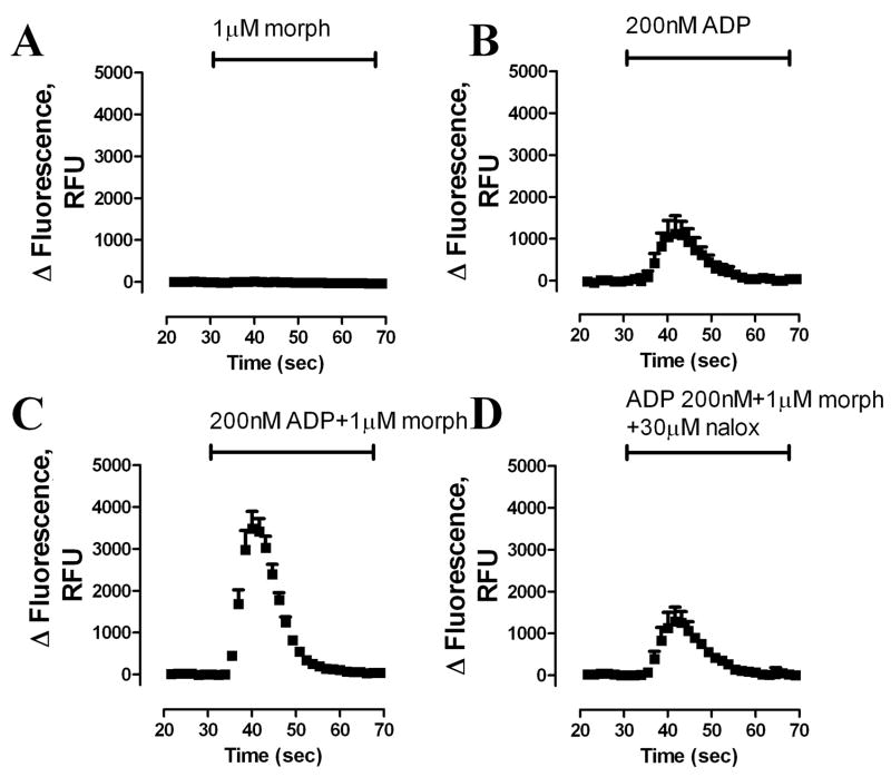
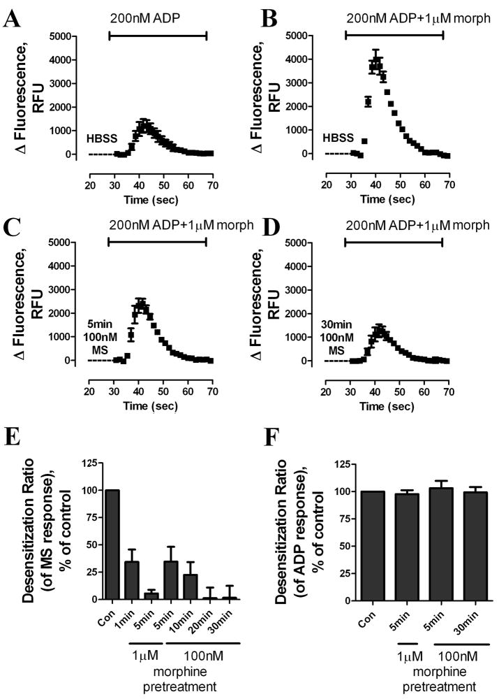
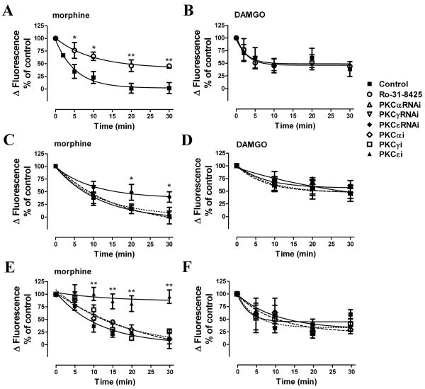
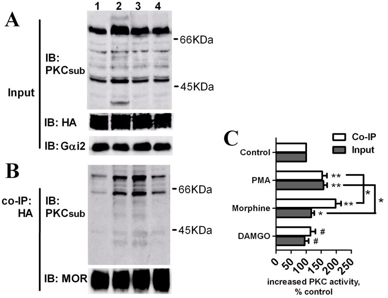
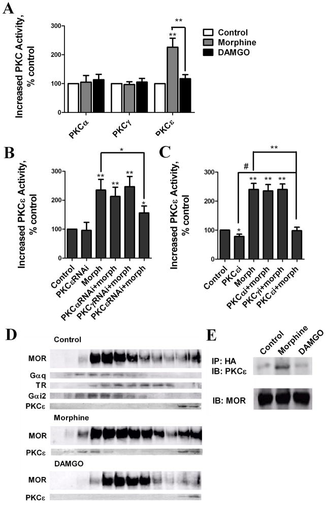
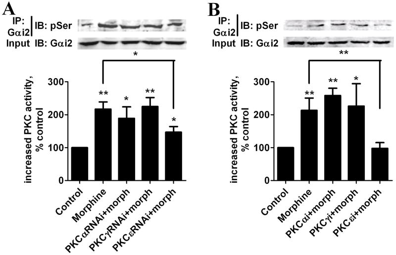
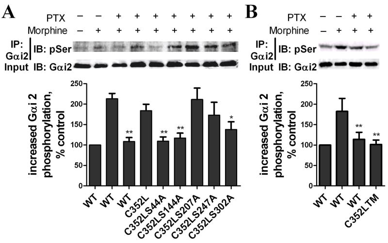
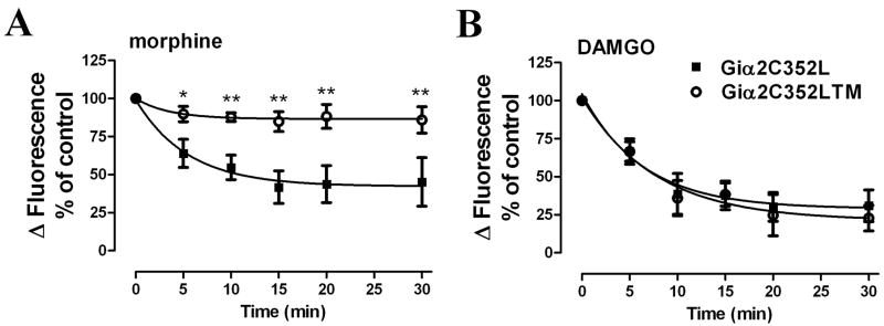
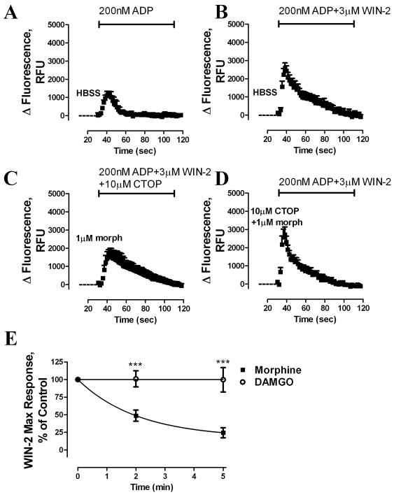
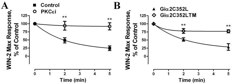
Desensitization of the micro-opioid receptor (MOR) has been implicated as an important regulatory process in the development of tolerance to opiates. Monitoring the release of intracellular Ca(2+) ([Ca(2+)](i)), we reported that [D-Ala(2), N-Me-Phe(4), Gly(5)-ol]-enkephalin (DAMGO)-induced receptor desensitization requires receptor phosphorylation and recruitment of beta-arrestins (betaArrs), while morphine-induced receptor desensitization does not. In current studies, we established that morphine-induced MOR desensitization is protein kinase C (PKC)-dependent. By using RNA interference techniques and subtype specific inhibitors, PKCepsilon was shown to be the PKC subtype activated by morphine and the subtype responsible for morphine-induced desensitization. In contrast, DAMGO did not increase PKCepsilon activity and DAMGO-induced MOR desensitization was not affected by modulating PKCepsilon activity. Among the various proteins within the receptor signaling complex, Galphai2 was phosphorylated by morphine-activated PKCepsilon. Moreover, mutating three putative PKC phosphorylation sites, Ser(44), Ser(144) and Ser(302) on Galphai2 to Ala attenuated morphine-induced, but not DAMGO-induced desensitization. In addition, pretreatment with morphine desensitized cannabinoid receptor CB1 agonist WIN 55212-2-induced [Ca(2+)](i) release, and this desensitization could be reversed by pretreating the cells with PKCepsilon inhibitor or overexpressing Galphai2 with the putative PKC phosphorylation sites mutated. Thus, depending on the agonist, activation of MOR could lead to heterologous desensitization and probable crosstalk between MOR and other Galphai-coupled receptors, such as the CB1.


Figures

Fig. 1
Fig. 1. MOR-potentiates Gq-coupled receptor-induced [Ca2+]i release
Real-time changes in intracellular fluorescence expressed in raw fluorescence units (RFU) were used to assess [Ca2+]i release from HA-tagged HEK293-MOR cells. Fluorescence changes were recorded using a 485-nm excitation wavelength and a 525-nm emission wavelength. After a 30-s baseline reading, agonists in HBSS buffer were added. (A) 1 μM morphine (morph); (B) 200 nM ADP; (C) 200 nM ADP + 1 μM morphine; (D) 200 nM ADP + 1 μM morphine + 30 μM naloxone (nalox) (n = 3).
Fig. 2
Fig. 2. Determining MOR desensitization by monitoring MOR-induced [Ca2+]i release
(A–D) Real-time changes in intracellular fluorescence are expressed in RFU. HEK293-MOR cells were pretreated with HBSS or 100 nM morphine for (C) 5 min or (D) 30 min, followed by ADP and additional morphine to achieve final agonists’ concentrations as indicated in the figure. (E) Dose response and time course of morphine-induced receptor desensitization. HEK293-MOR cells were treated as described in (A–D). Total [Ca2+]i release after pretreatment was quantified by calculating the area under curves with a Prism program. Response to ADP was subtracted from total response to obtain the response to MOR. MOR response after morphine pretreatment was compared to MOR response after HBSS pretreatment to obtain the desensitization ratio. (F) Effect of morphine pretreatment on response to 1 μM ADP. After morphine pretreatment, ADP was added together with 10μM CTOP to block MOR response. Response to ADP was analyzed as described in Fig. 2E. (n≥3)
Fig. 3
Fig. 3. Morphine-induced, but not DAMGO-induced, MOR desensitization is PKC-dependent
Morphine-induced desensitization as measured by assessing agonist potentiation of ADP-mediated [Ca2+]i release. 100 nM morphine or DAMGO were used to pretreat HEK293-MOR cells for various time indicated in the X-axis. Desensitization ratios were determined as described in Fig. 2. HEK293-MOR cells were incubated for 3 h with DMSO (Control) (■), 5 μM general PKC inhibitor Ro-31-8425 (○), transfected for 48 h with PKCαRNAi (△), PKCγRNAi (▽), PKCεRNAi (◆) or incubated for 3 h 50 μM subtype-specific inhibitors of PKCα (PKCαi) (◇), γ (PKCγi) (□), or ε (PKCεi) (▲) before assays (*: p < 0.05; **: p < 0.01; n≥3).
Fig. 4
Fig. 4. Activation of PKC by morphine treatment
HEK293-MOR cells were pretreated with Buffer (lane 1), 1μM PMA (lane 2), 1μM morphine (lane 3) or 1μM DAMGO (lane 4) for 5 mins. (A) PKC activity in whole cell lysates was determined by immunoblotting (IB) with an antibody against phosphorylated PKC substrates (PKCsub), and Gαi2 and MOR in whole cell lysates were used as input controls. (B) MOR signaling complexes were co-immumoprecipitated (co-IP) with HA antibody, and PKC activity was determined by IB with PKCsub; MOR within immumoprecipitates was determined with an antibody against MOR C-terminal. (C) Quantitative analysis of immunoreactivities of PKC phosphorylated substrates as determined from IBs. (#: no significant difference; *: p < 0.05; **: p < 0.01; n=3).
Fig. 5
Fig. 5. Morphine increases PKCε activity and translocates PKCε to the MOR signaling complex
(A) Determination of agonist-induced PKC subtype activity in cell lysates. HEK293-MOR cells were pretreated for 5 min with 1 μM morphine and DAMGO. Activities of individual PKC subtypes were determined (*: p < 0.05; **: p < 0.01; n>3). (B) Determination of morphine-induced PKCε activity in cell lysates after overexpression of PKC subtypes RNAis. HEK293-MOR cells were transfected with PKCαRNAi, PKCγRNAi or PKCεRNAi for 48 h before 5 min 1μM morphine pretreatment. Activities of PKCε subtype were determined (*: p < 0.05; **: p < 0.01; n≥3) (C) Determination of morphine-induced PKCε activity in cell lysates after. HEK293-MOR cells were incubated for 3 h 50 μM PKCε subtype-specific inhibitor (PKCεi) before 5 min 1μM morphine pretreatment. Activities of PKCε subtype were determined (*: p < 0.05; **: p < 0.01; n≥3) (D) Morphine-induced translocation of PKCε to the MOR signaling complex. Cells were pretreated with 1 μM morphine or DAMGO for 5 mins. Homogenates were fractionated on continuous sucrose gradients. Gαi2 and Gq were used as lipid raft markers; transferrin receptor (TR) as a non-raft marker Lanes. Left to right: sucrose gradient fractions 1–12. (E) Morphine-induced PKCε association with the MOR signaling complex. Cells were pretreated with 1 μM morphine or DAMGO for 5 min. MOR signaling complexes were immunoprecipitated with HA antibody and immunoblotted with PKCε antibody; MOR was used as loading control.
Fig. 6
Fig. 6. Gαi2 is essential for MOR signaling transduction
(A–D) PTX-resistant Gαi2 mutants Gαi2C352L (A), Gαi3C351L (B), GαoC352L (C) or wild type Gαi2 (Gαi2WT; D) were overexpressed in HEK293-MOR cells for 48 h. Cells were pretreated with HBSS (Con) (■), PTX 20ng/ml (○) or PTX 100ng/ml (▲) for 16 h before morphine-induced [Ca2+]i release dose-response assays. (E) Vector (□), Gαi2 sense (S) (●) or antisense (AS) (▼;) constructs were overexpressed for 48 h before morphine-induced [Ca2+]i release dose-response assays. Different concentrations of morphine were added with 200nM ADP. Total [Ca2+]i release after pretreatment was quantified by calculating the area under curves with a Prism program. Response to ADP was subtracted from total response to obtain the response to morphine (*: p < 0.05; **: p < 0.01; n=3).
Fig. 7
Fig. 7. Gαi2 is phosphorylated by PKCε after morphine treatment
HEK293-MOR cells were transfected with PKCαRNAi, PKCγRANi or PKCεRNAi for 48 h (A) or preincubated with specific inhibitors of PKC subtypes α (PKCαi), γ (PKCγi)or ε (PKCεi)for 3 h (B) before assays. Cells were pretreated with 1 μM morphine. Gαi2 was immunoprecipitated, and Gαi2 phosphorylation was determined using pSer antibody. Gαi2 in whole cell lysates was used as a loading control. (top): immunoblots; (bottom): quantitative analysis of immunoblots (#: no significant difference; *: p < 0.05; **: p < 0.01; n=3).
Fig. 8
Fig. 8. Identifying morphine-induced Gαi2 phosphorylation sites
(A) HEK293-MOR cells were transfected for 48 h with wild type Gαi2 plasmid (WT) PTX-resistant (C352L) Gαi2 plasmid or C352L constructs mutated at serine residues 44, 144, 207, 247 or 302 to alanine. Cells were pretreated with 100 ng/ml PTX for 16 h and with 1 μM morphine for 5 min before determination of Gαi2 phosphorylation as described in legend of Fig. 7. (B) Cells transfected with wild type Gαi2 plasmid (WT) or C352L mutated at serine residues 44, 144, and 302 (triple mutation; C352LTM) Phosphorylation of the Gαi2 was determined as described previously (*: p < 0.05; **: p < 0.01; n=3).
Fig. 9
Fig. 9. Morphine-induced MOR desensitization is mediated by PKC-induced Gαi2 phosphorylation
MOR desensitization was determined by pretreating cells with (A) 100 nM morphine or (B) 100 nM DAMGO for various time as indicated in the X-axis. HEK293-MOR cells were transfected with Gαi2C352L (■) or Gαi2C352LTM (○) for 48 h and pretreated with 100 ng/ml PTX for 16 h before the desensitization assays (*: p < 0.05; **: p < 0.01; n=3).
Fig. 10
Fig. 10. Morphine, but not DAMGO, induces heterologous desensitization of CB1 receptor
Time-dependent changes in intracellular fluorescence (RFU) were used to assess [Ca2+]i release. HEK293-MOR cells transiently expressing the CB1 receptor were pretreated with (A–B) HBSS, (C) 1 μM morphine, or (D) 1 μM morphine + 10 μM CTOP for 5 min, followed addition of (A) 200 nM ADP, (B, D) 200 nM ADP + 3 μM Win-2, or (C) 200 nM ADP + 3 μM Win-2 + 10 μM CTOP. (E) CB1 desensitization after pretreatment with 1 μM morphine or DAMGO was measured at the time indicated in the X-axis (***: p < 0.001; n=3).
Fig. 11
Fig. 11. Morphine-induced CB1 heterologous desensitization is mediated by PKCε-induced Gαi2 phosphorylation
(A) Morphine-induced heterologous CB1 desensitization is PKCε dependent. Heterologous desensitization assays were performed as described in Fig. 10 After morphine pretreatment, the Win-2 induced maximum response was compared to the control. Cells were pretreated with PKCε inhibitor (PKCεi) (○) or DMSO (Control) (■) for 3 h before assays. (B) Morphine-induced heterologous CB1 desensitization is Gαi2-phosphorylation-dependent. HEK293-MOR cells transiently expressing the CB1 receptor overexpressing Gαi2C352L or Gαi2C352LTM was pretreated with 100 ng/ml PTX for 16 h before the desensitzation assays (**: p < 0.01; n=3).

Similar articles

Cited by

References

    1. Borgland SL. Clin Exp Pharmacol Physiol. 2001;28(3):147–154. - PubMed
    1. Lefkowitz RJ. J Biol Chem. 1998;273(30):18677–18680. - PubMed
    1. Violin JD, Lefkowitz RJ. Trends Pharmacol Sci. 2007;28(8):416–422. - PubMed
    1. Gainetdinov RR, Premont RT, Bohn LM, Lefkowitz RJ, Caron MG. Annu Rev Neurosci. 2004;27:107–144. - PubMed
    1. Chu J, Zheng H, Loh HH, Law PY. Cell Signal. 2008;20(9):1616–1624. - PMC - PubMed

Publication types

MeSH terms

Substances