Brain injury and recovery following binge ethanol: evidence from in vivo magnetic resonance spectroscopy
- PMID: 20044076
- PMCID: PMC2854208
- DOI: 10.1016/j.biopsych.2009.10.028
Brain injury and recovery following binge ethanol: evidence from in vivo magnetic resonance spectroscopy
Abstract
Background: The binge-drinking model in rodents using intragastric injections of ethanol (EtOH) for 4 days results in argyrophilic corticolimbic tissue classically interpreted as indicating irreversible neuronal degeneration. However, recent findings suggest that acquired argyrophilia can also identify injured neurons that have the potential to recover. The current in vivo magnetic resonance (MR) imaging and spectroscopy study was conducted to test the hypothesis that binge EtOH exposure would injure but not cause the death of neurons as previously ascertained postmortem.
Methods: After baseline MR scanning, 11 of 19 rats received a loading dose of 5 g/kg EtOH via oral gavage, then a maximum of 3 g/kg every 8 hours for 4 days, for a total average cumulative EtOH dose of 43 +/- 1.2 g/kg and average blood alcohol levels of 258 +/- 12 mg/dL. All animals were scanned after 4 days of gavage (post-gavage scan) with EtOH (EtOH group) or dextrose (control [Con] group) and again after 7 days of abstinence from EtOH (recovery scan).
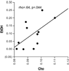
Results: Tissue shrinkage at the post-gavage scan was reflected by significantly increased lateral ventricular volume in the EtOH group compared with the Con group. At the post-gavage scan, the EtOH group had lower dorsal hippocampal N-acetylaspartate and total creatine and higher choline-containing compounds than the Con group. At the recovery scan, neither ventricular volume nor metabolite levels differentiated the groups.
Conclusions: Rapid recovery of ventricular volume and metabolite levels with removal of the causative agent argues for transient rather than permanent effects of a single EtOH binge episode in rats.
Copyright 2010 Society of Biological Psychiatry. Published by Elsevier Inc. All rights reserved.
Conflict of interest statement
The authors report no biomedical financial interests or potential conflicts of interest.
Figures


References
-
- Sullivan EV, Pfefferbaum A. Neurocircuitry in alcoholism: A substrate of disruption and repair. Psychopharmacology (Berl) 2005;180:583–594. - PubMed
-
- Sullivan EV. Compromised pontocerebellar and cerebellothalamocortical systems: speculations on their contributions to cognitive and motor impairment in nonamnesic alcoholism. Alcoholism, clinical and experimental research. 2003;27:1409–1419. - PubMed
-
- Pfefferbaum A, Sullivan EV, Mathalon DH, Shear PK, Rosenbloom MJ, Lim KO. Longitudinal changes in magnetic resonance imaging brain volumes in abstinent and relapsed alcoholics. Alcoholism: Clinical and Experimental Research. 1995;19:1177–1191. - PubMed
Publication types
MeSH terms
Substances
Grants and funding
LinkOut - more resources
Full Text Sources
Medical

