. 2010 Apr;38(6):2044-56.
doi: 10.1093/nar/gkp1187. Epub 2009 Dec 30.

Control of directionality in the DNA strand-exchange reaction catalysed by the tyrosine recombinase TnpI

Affiliations

Control of directionality in the DNA strand-exchange reaction catalysed by the tyrosine recombinase TnpI

Virginie Vanhooff et al. Nucleic Acids Res. 2010 Apr.

Abstract

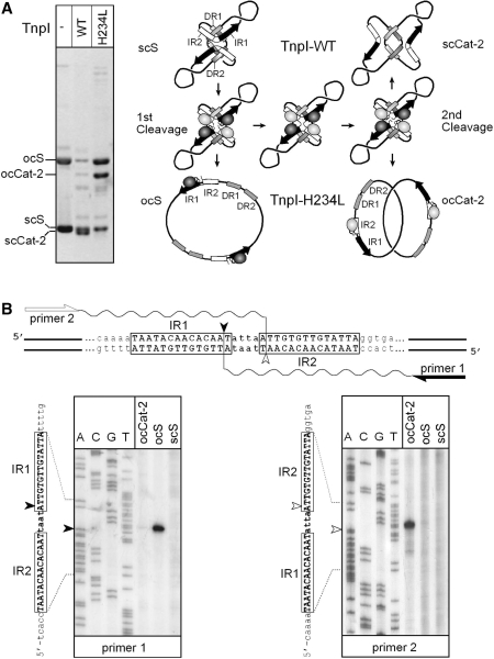
In DNA site-specific recombination catalysed by tyrosine recombinases, two pairs of DNA strands are sequentially exchanged between separate duplexes and the mechanisms that confer directionality to this theoretically reversible reaction remain unclear. The tyrosine recombinase TnpI acts at the internal resolution site (IRS) of the transposon Tn4430 to resolve intermolecular transposition products. Recombination is catalysed at the IRS core sites (IR1-IR2) and is regulated by adjacent TnpI-binding motifs (DR1 and DR2). These are dispensable accessory sequences that confer resolution selectivity to the reaction by stimulating synapsis between directly repeated IRSs. Here, we show that formation of the DR1-DR2-containing synapse imposes a specific order of activation of the TnpI catalytic subunits in the complex so that the IR1-bound subunits catalyse the first strand exchange and the IR2-bound subunits the second strand exchange. This ordered pathway was demonstrated for a complete recombination reaction using a TnpI catalytic mutant (TnpI-H234L) partially defective in DNA rejoining. The presence of the DR1- and DR2-bound TnpI subunits was also found to stabilize transient recombination intermediates, further displacing the reaction equilibrium towards product formation. Implication of TnpI/IRS accessory elements in the initial architecture of the synapse and subsequent conformational changes taking place during strand exchange is discussed.


Figures

Figure 1.
Figure 1.
The strand-exchange reaction catalysed by tyrosine recombinases. Binding of a pair of recombinase molecules (purple ovals) onto the core recombination site recognition elements (open arrows) may bend DNA to opposite directions (a or b) exposing one or the other strand for catalysis (coloured dark and light green, respectively). In all cases studied, activity of the recombinase subunits is controlled by allosteric interactions in which the active subunit donates its C-terminus (represented by a stem-and-ball extension) to its neighbour. During synaptic complex formation (I), both recombination sites may pair with an antiparallel alignment if they are in the same bent configuration (a × a or b × b) or with a parallel alignment if they are bent in opposite directions (a × b or b × a). In all cases, this leads to the formation of a recombinase tetramer in which diagonally opposed subunits are activated, while the other pair of subunits is inactive. Antiparallel pairing of the recombination sites proceeds to strand exchange through a sequence of reversible steps during which the exposed strands are cleaved (II), exchanged (III) and then rejoined (IV) to the partner strand to form a HJ. Isomerization of the HJ (V) inactivates one pair of recombinase subunits and activates the other pair of subunits to exchange the second pair of strands. For each strand-exchange reaction, the recombinase catalytic tyrosine (purple arrowhead) attacks the adjacent sessile phosphate (yellow circle) to form a phosphotyrosyl bond that is in turn attacked by the 5′OH end of the cleaved DNA strand (green arrowhead). Note that recombination that initiates with one configuration of the synapse terminates with the core sites in the opposite configuration and reciprocally. Parallel pairing of the recombination sites generally leads to abortive recombination due to the lack base pairs complementarity (red crosses) in the overlap region (step III).
Figure 2.
Figure 2.
The TnpI/IRS resolution system of Tn4430. (A) Structure of the IRS recombination site and its derivatives. Wild-type IRS (IRS1.2, 116 bp) contains two 14-bp inverted motifs, IR1 (filled triangle) and IR2 (open triangle), forming the recombination core site; and two 16 bp accessory TnpI-binding motifs, DR1 and DR2 (shaded triangles), playing a regulatory role in recombination. All motifs share a common sequence of 9 bp (boxed) thought to be bound by separate recombinase molecules within the recombination complex. The distance that separates the different motifs (in bp) is indicated. TnpI-mediated DNA cleavages at IR1 and IR2 take place at symmetrical positions staggered by 6 bp as indicated by a filled and open arrowhead, respectively. Both motifs are separated by a spacer sequence of 4 bp (orientation shown by an arrow). The IRS2.1 variant was constructed by reversing the orientation of the IR1–IR2 core site (Core1.2) with respected to the DR1–DR2 accessory motifs. IRS1.1 and IRS2.2 were constructed by replacing the IR1–IR2 core site with symmetrical sequences containing inversely oriented IR1 (Core1.1) and IR2 (Core2.2) half sites, respectively. Simple core recombination sites corresponding to the different IRS variants were made by replacing the DR1–DR2 sequence with an unrelated DNA fragment of the same length (see the ‘Materials and Methods’ section). (B) Topologically constrained and relaxed recombination activity of TnpI. TnpI-mediated recombination of supercoiled DNA molecules carrying directly repeated full-length IRSs exclusively generates two-noded catenane products (2-Cat) following the formation of a topologically defined synaptic complex involving the accessory motifs DR1 and DR2 (I). In the absence of DR1-DR1, recombination between minimal IR1–IR2 core sites takes place after random collision, trapping a variable number of DNA supercoils between both recombination sites and generating a mixture of catenanes (x-Cat) containing an even number of crossings (or nodes) if the two sites are in direct repeat (II), or a mixture of unknotted and knotted inversion products (x-Knt) containing an odd number of nodes if the two sites are in inverted repeat (III).
Figure 3.
Figure 3.
TnpI-mediated relaxed and constrained recombination between directly and inversely repeated core sites in vivo. Reporter plasmids containing different arrangements of the IR1–IR2 core sites joined to directly repeated DR1–DR2 sequences or not were transformed into E. coli TOP10 cells harbouring the TnpI expression vector pGIV004 (ev) or the control plasmid pCB104 (c). Plasmid DNA was isolated from a pool of transformants and run uncut on a 0.8% agarose gel. Intramolecular recombination of the substrate monomer (S) yielded monomeric resolution (mR) products in which the DNA fragment between both recombination sites was deleted. Unconstrained recombination generated multimers of the substrate and/or resolution product, together with a mixture of high molecular weight products arising from both inter and intramolecular recombination reactions. Reporter plasmids are diagrammed on the top of the figure, with the IR1 and IR2 core motifs represented by filled and open triangles, respectively; and the DR1 and DR2 accessory motifs by shaded triangles. Arrows show the orientation of the core sites as determined by the central spacer sequence.
Figure 4.
Figure 4.
TnpI-mediated recombination at the reversed core site IRS2.1 variant in vitro. Supercoiled plasmids carrying directly repeated copies of wild-type IRS (IRS1.2 × 1.2) or modified IRS in which the orientation of the IR1–IR2 core site is reversed with respect to the DR1–DR2 accessory sequence (IR2.1 × 2.1) were incubated for the indicated times with purified TnpI. Reaction were digested with AvaII (AII) and analysed by electrophoresis on a 0.8% agarose gel. S1, S2 and S3 are the AvaII fragments from the initial substrates. D1 and D2 are specific fragments from the deletion products. AvaII digestion of the reactions also produced χ-forms corresponding to the Holliday junction (HJ) intermediate of recombination.
Figure 5.
Figure 5.
TnpI-mediated recombination at symmetrical core sites in vitro. Reporter plasmids carrying tandem copies of the wild-type (Core1.2) or symmetrical (Core1.1 and Core2.2) core sites under the control of DR1–DR2 accessory sequences (panels IV–VI) or not (panels I–III) were incubated with purified TnpI for the indicated times. Reactions were digested with SmaI (SI) and analysed by agarose gel electrophoresis. The cartoon summarizes all possible DNA fragments that may arise from recombination at both substrate sites (site1 and 2) after digestion with SmaI. S1 and S2 are the initial substrate fragments. D1 and D2 are DNA fragments arising from recombination between directly oriented sites1 and 2. Inv1 and Inv2 are recombination products of inversely oriented sites. Inter1, Inter2, Inter3 and Inter4 are specific ‘intermolecular’ site1 × site1 and site2 × site2 recombination products in which flanking DNA segments are joined in head-to-head and tail-to-tail configuration (Inter4 was too small to be seen on the gel). HJ intermediates migrate as a χ-form at an upper position of the gel.
Figure 6.
Figure 6.
DNA topology of recombination products. Recombination of circular substrates containing pairs of core site variants in the absence (panels I–III) or presence (panels IV–VII) of directly repeated DR1–DR2 accessory motifs were treated with TnpI or left untreated as indicated. Reactions were singly nicked with DNase I and run on a 0.8% agarose gel. Positions of the open circular (ocS), linear (lin), and supercoiled (scS) forms of the substrates are indicated, as are the positions of the nicked two-, three-, four-, five-, six-, seven- and eight-noded catenane and knot products of recombination. Note that the majority of secondary (twist) knots generated by consecutive rounds of recombination at Core1.2 (panel I) migrate slightly ahead of the primary (torus) knots arising from recombination at the symmetrical Core1.1 and Core2.2 sites (panels II and III). Recombination of the DR1 and DR2-containg plasmids (panels IV–VII) yielded two-noded catenanes (ocCat-2) as the only product of recombination. Additional bands (asterisk) correspond to circular deletion products that were released from catenanes by over digestion with DNase I.
Figure 7.
Figure 7.
Analysis of HJ intermediates formed at different variants of IRS. HJ products that accumulated in recombination reactions performed with different IRS substrates (see the ‘Materials and Methods’ section) were cut with AvaII (Av), gel purified, and 5′-labelled with 32P (asterisk). The resulting χ-forms were left untreated (−) or recut with NruI (Nr) or ScaI (Sc) and run on DNA denaturing gels. (A) Different patterns of radio-labelled single-stranded DNA fragments are expected depending on whether the HJ is formed by the IR1-bound TnpI subunits (1 × 1′), the IR2-bound subunits (2 × 2′) or opposite combinations of IR1- and IR2-bound subunits (1 × 2′ and 2 × 1′, respectively). Sizes (in nt) of the diagnostic fragments obtained for the different possibilities of strand exchange are reported in the corresponding tables. One fragment (in bold) is specific for each possibility and the corresponding DNA strand is shown as a thick line in the cartoons showing the different HJ isoforms. (B) Bands patterns obtained for the different substrates after autoradiography of the gels. For each group of substrates, sizes of the initial χ-form fragments are indicated on the left, and the new fragments that were generated after NruI and ScaI cleavage are shown on the right. All the DR1–DR2-constraining substrates (panels I–V) show a unique pattern of bands corresponding to the 1 × 1′ exchange, irrespective of the nature and orientation of the core sites. The pattern obtained for the pCore1.2 × 1.2 substrate (panel VI) is consistent with strand exchange occurring at both IR1 and IR2. HJ formation at the symmetrical Core1.1 and Core2.2 sites (panel VII and VIII) occurred with all possibilities of strand exchange.
Figure 8.
Figure 8.
Ordered DNA cleavages during TnpI-catalysed sequential strand exchanges (A) TnpI and TnpI-H234L recombination pathways. A reporter plasmid carrying directly repeated IRSs was incubated with wild-type TnpI (TnpI-WT) or TnpI-H234L, and the reactions were run uncut on a 0.8% agarose gel. Both proteins assembled onto the supercoiled substrate (scS) to form the TnpI/IRS-specific synapse. For clarity, only one of both possible topological organizations of the synaptic complex is shown (19). The IR1–IR2 core sites and the DR1 and DR2 accessory motifs are represented by black/white arrows and shaded boxes, respectively. The core TnpI subunits bound to IR1 and IR2 half sites are shown as dark and light shaded ovals, respectively. Sequential strand-exchange reactions catalysed by TnpI-WT leads to fully supercoiled two-noded catenane products (scCat-2). In the presence of TnpI-H234L, DNA rejoining was partially inhibited leading to the formation of open circular forms of the substrate (ocS) and catenane product (ocCat-2). (B) Mapping of TnpI-H234L DNA cleavages. Gel-purified products arising from the first (ocS) and second (ocCat-2) DNA cleavage reactions were used as a template in primer extension analysis to determine whether cleavage occurred at IR1 (primer 1) or IR2 (primer 2). Extension products were analysed on a 6% sequencing gel alongside of a DNA sequencing reaction performed with the same primers. Extensions performed with unreacted substrate (scS) were used as a control in the experiment. IR1 and IR2 cleavage positions are shown by a filled and open arrowhead, respectively. Note that the thermostable DNA polymerase used in the experiment added an extra dA nucleotide at the 3′ end of the extension products.

Similar articles

Cited by

References

    1. Craig N, Craigie R, Gellert M, Lambowitz A, editors. Mobile DNA II. Washington, DC: ASM Press; 2002.
    1. Hallet B, Sherratt DJ. Transposition and site-specific recombination: adapting DNA cut-and-paste mechanisms to a variety of genetic rearrangements. FEMS Microbiol. Rev. 1997;21:157–178. - PubMed
    1. Curcio MJ, Derbyshire KM. The outs and ins of transposition: from mu to kangaroo. Nat. Rev. Mol. Cell Biol. 2003;4:865–877. - PubMed
    1. Van Duyne GD. A structural view of cre-loxp site-specific recombination. Annu. Rev. Biophys. Biomol. Struct. 2001;30:87–104. - PubMed
    1. Grindley ND, Whiteson KL, Rice PA. Mechanisms of site-specific recombination. Annu. Rev. Biochem. 2006;75:567–605. - PubMed

Publication types