

Signaling responses after exposure to 5 alpha-dihydrotestosterone or 17 beta-estradiol in norepinephrine-induced hypertrophy of neonatal rat ventricular myocytes
- PMID: 20044473
- PMCID: PMC2838630
- DOI: 10.1152/japplphysiol.00994.2009
Signaling responses after exposure to 5 alpha-dihydrotestosterone or 17 beta-estradiol in norepinephrine-induced hypertrophy of neonatal rat ventricular myocytes
Abstract
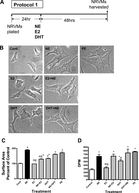
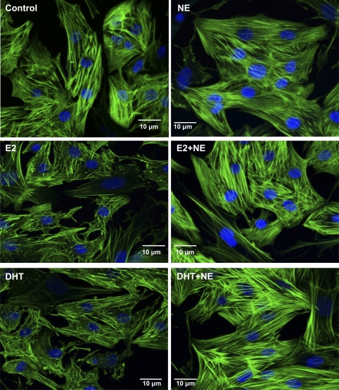
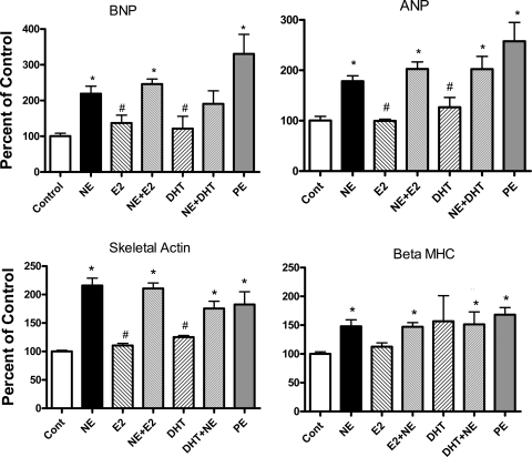
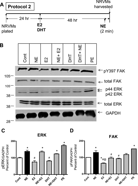
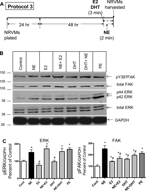
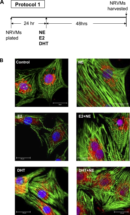
Androgens appear to enhance, whereas estrogens mitigate, cardiac hypertrophy. However, signaling pathways in cells for short (3 min) and longer term (48 h) treatment with 17beta-estradiol (E2) or 5 alpha-dihydrotestosterone (DHT) are understudied. We compared the effect of adrenergic stimulation by norepinephrine (NE; 1 microM) alone or in combination with DHT (10 nM) or E2 (10 nM) treatment in neonatal rat ventricular myocytes (NRVMs) by cell area, protein synthesis, sarcomeric structure, gene expression, phosphorylation of extracellular signal-regulated (ERK), and focal adhesion kinases (FAK), and phospho-FAK nuclear localization. NE alone elicited the expected hypertrophy and strong sarcomeric organization, and DHT alone gave a similar but more modest response, whereas E2 did not alter cell size. Effects of NE dominated when used with either E2 or DHT with all combinations. Both sex hormones alone rapidly activated FAK but not ERK. Long-term or brief exposure to E2 attenuated NE-induced FAK phosphorylation, whereas DHT had no effect. Neither hormone altered NE-elicited ERK activation. Longer term exposure to E2 alone reduced FAK phosphorylation and reduced nuclear phospho-FAK, whereas its elevation was seen in the presence of NE with both sex hormones. The mitigating effects of E2 on the NE-elicited increase in cell size and the hypertrophic effect of DHT in NRVMs are in accordance with results observed in whole animal models. This is the first report of rapid, nongenomic sex hormone signaling via FAK activation and altered FAK trafficking to the nucleus in heart cells.
Figures
Similar articles
-
Shp2 negatively regulates growth in cardiomyocytes by controlling focal adhesion kinase/Src and mTOR pathways.Circ Res. 2008 Oct 10;103(8):813-24. doi: 10.1161/CIRCRESAHA.108.179754. Epub 2008 Aug 28. Circ Res. 2008. PMID: 18757826
-
Focal adhesion kinase and p130Cas mediate both sarcomeric organization and activation of genes associated with cardiac myocyte hypertrophy.Mol Biol Cell. 2001 Aug;12(8):2290-307. doi: 10.1091/mbc.12.8.2290. Mol Biol Cell. 2001. PMID: 11514617 Free PMC article.
-
Endothelin-induced cardiac myocyte hypertrophy: role for focal adhesion kinase.Am J Physiol Heart Circ Physiol. 2000 May;278(5):H1695-707. doi: 10.1152/ajpheart.2000.278.5.H1695. Am J Physiol Heart Circ Physiol. 2000. PMID: 10775151
-
ErbB/integrin signaling interactions in regulation of myocardial cell-cell and cell-matrix interactions.Biochim Biophys Acta. 2013 Apr;1833(4):909-16. doi: 10.1016/j.bbamcr.2012.12.007. Epub 2012 Dec 20. Biochim Biophys Acta. 2013. PMID: 23261977 Free PMC article. Review.
-
Focal adhesion kinase -- the basis of local hypertrophic signaling domains.J Mol Cell Cardiol. 2012 Feb;52(2):485-92. doi: 10.1016/j.yjmcc.2011.06.021. Epub 2011 Jul 2. J Mol Cell Cardiol. 2012. PMID: 21749874 Review.
Cited by
-
What we know and do not know about sex and cardiac disease.J Biomed Biotechnol. 2010;2010:562051. doi: 10.1155/2010/562051. Epub 2010 Apr 22. J Biomed Biotechnol. 2010. PMID: 20445744 Free PMC article. Review.
-
Real-time monitoring of hypertrophy in HL-1 cardiomyocytes by impedance measurements reveals different modes of growth.Cytotechnology. 2016 Oct;68(5):1897-907. doi: 10.1007/s10616-016-0001-3. Epub 2016 Jul 5. Cytotechnology. 2016. PMID: 27380966 Free PMC article.
-
Mechanical stress-induced sarcomere assembly for cardiac muscle growth in length and width.J Mol Cell Cardiol. 2010 May;48(5):817-23. doi: 10.1016/j.yjmcc.2010.02.016. Epub 2010 Feb 24. J Mol Cell Cardiol. 2010. PMID: 20188736 Free PMC article. Review.
-
Striated muscle proteins are regulated both by mechanical deformation and by chemical post-translational modification.Biophys Rev. 2021 Sep 4;13(5):679-695. doi: 10.1007/s12551-021-00835-4. eCollection 2021 Oct. Biophys Rev. 2021. PMID: 34777614 Free PMC article. Review.
-
Overweight female rats selectively breed for low aerobic capacity exhibit increased myocardial fibrosis and diastolic dysfunction.Am J Physiol Heart Circ Physiol. 2012 Apr 15;302(8):H1667-82. doi: 10.1152/ajpheart.01027.2011. Epub 2012 Feb 17. Am J Physiol Heart Circ Physiol. 2012. PMID: 22345570 Free PMC article.
References
-
- Agabiti-Rosei E, Muiesan ML. Left ventricular hypertrophy and heart failure in women. J Hypertens Suppl 20: S34–S38, 2002 - PubMed
-
- Ahluwalia A, Clodfelter KH, Waxman DJ. Sexual dimorphism of rat liver gene expression: regulatory role of growth hormone revealed by deoxyribonucleic acid microarray analysis. Mol Endocrinol 18: 747–760, 2004 - PubMed
-
- Altamirano F, Oyarce C, Silva P, Toyos M, Wilson C, Lavandero S, Uhlen P, Estrada M. Testosterone induces cardiomyocyte hypertrophy through mammalian target of rapamycin complex 1 pathway. J Endocrinol 202: 299–307, 2009 - PubMed
-
- Babiker FA, De Windt LJ, van Eickels M, Thijssen V, Bronsaer RJ, Grohe C, van Bilsen M, Doevendans PA. 17beta-estradiol antagonizes cardiomyocyte hypertrophy by autocrine/paracrine stimulation of a guanylyl cyclase A receptor-cyclic guanosine monophosphate-dependent protein kinase pathway. Circulation 109: 269–276, 2004 - PubMed
-
- Barki-Harrington L, Perrino C, Rockman HA. Network integration of the adrenergic system in cardiac hypertrophy. Cardiovasc Res 63: 391–402, 2004 - PubMed
Publication types
MeSH terms
Substances
Grants and funding
LinkOut - more resources
Full Text Sources
Miscellaneous