. 2010 Mar 5;285(10):7067-78.
doi: 10.1074/jbc.M109.006841. Epub 2009 Dec 31.

Intracellular calreticulin regulates multiple steps in fibrillar collagen expression, trafficking, and processing into the extracellular matrix

Affiliations

Intracellular calreticulin regulates multiple steps in fibrillar collagen expression, trafficking, and processing into the extracellular matrix

Lauren Van Duyn Graham et al. J Biol Chem. .

Abstract

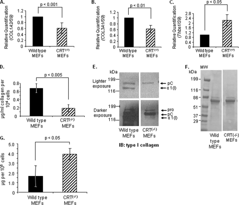
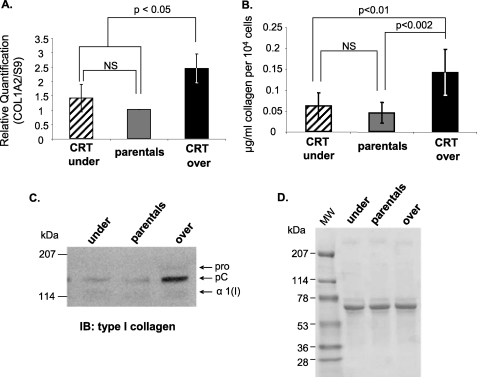
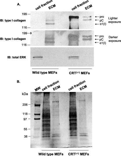
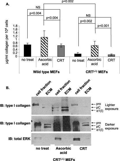
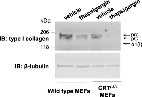
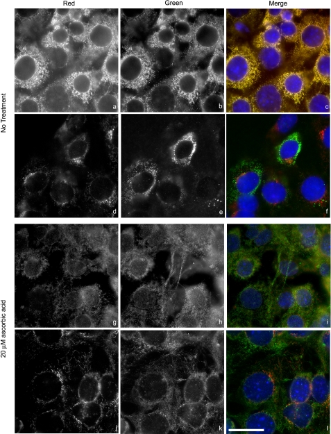
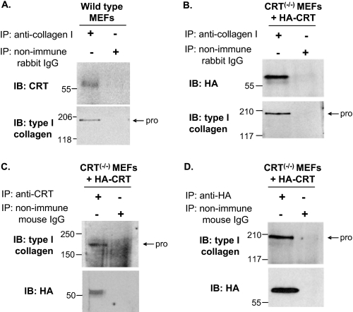
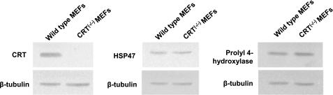
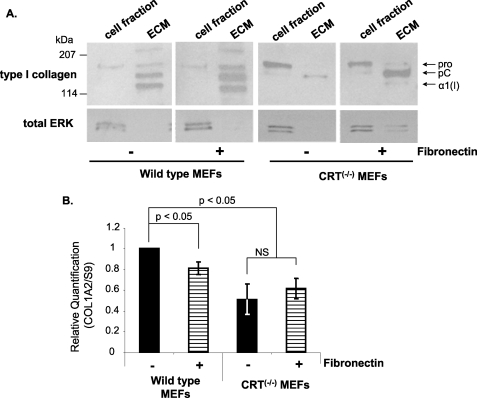
Calreticulin (CRT), a chaperone and Ca(2+) regulator, enhances wound healing, and its expression correlates with fibrosis in animal models, suggesting that CRT regulates production of the extracellular matrix. However, direct regulation of collagen matrix by CRT has not been previously demonstrated. We investigated the role of CRT in the regulation of fibrillar collagen expression, secretion, processing, and deposition in the extracellular matrix by fibroblasts. Mouse embryonic fibroblasts deficient in CRT (CRT(-/-) MEFs) have reduced transcript levels of fibrillar collagen I and III and less soluble collagen as compared with wild type MEFs. Correspondingly, fibroblasts engineered to overexpress CRT have increased collagen type I transcript and protein. Collagen expression appears to be regulated by endoplasmic reticulum (ER) calcium levels and intracellular CRT, because thapsigargin treatment reduced collagen expression, whereas addition of exogenous recombinant CRT had no effect. CRT(-/-) MEFs exhibited increased ER retention of collagen, and collagen and CRT were co-immunoprecipitated from isolated cell lysates, suggesting that CRT is important for trafficking of collagen through the ER. CRT(-/-) MEFs also have reduced type I procollagen processing and deposition into the extracellular matrix. The reduced collagen matrix deposition is partly a consequence of reduced fibronectin matrix formation in the CRT-deficient cells. Together, these data show that CRT complexes with collagen in cells and that CRT plays critical roles at multiple stages of collagen expression and processing. These data identify CRT as an important regulator of collagen and suggest that intracellular CRT signaling plays an important role in tissue remodeling and fibrosis.


Figures

FIGURE 1.
FIGURE 1.
Cells deficient in CRT expression have reduced collagen transcript and soluble collagen. A–C, wild type and CRT−/− MEFs were cultured in 10% FBS for 48 h and then 12 h in 0.5% FBS. RNA was harvested, and transcript levels of type I collagen, Col1A2 (A), type III collagen, Col3A1 (B), and Thbs1 (C) were measured by quantitative real time PCR. Values represent the mean expression levels normalized to S9 expression ± S.D. (for Col1A2, n = 8; for Col3A1, n = 5; for Thbs1, n = 4; each “n” was performed in triplicate). Values for wild type cells were set to 1. D–F, wild type and CRT−/− MEFs were cultured in 10% FBS for 48 h and then 72 h in 0.5% FBS. Conditioned media were collected after the 72 h. D, conditioned media were assayed for levels of soluble collagen using the SircolTM assay. Soluble collagen levels were normalized to cell number. Results are expressed as the mean ± S.D. (n = 3 separate experiments performed in triplicate). E, equal volumes of conditioned media were immunoblotted (IB) for type I collagen. A representative blot is shown with a lighter exposure to visualize collagen in the wild type MEFs and a darker exposure to visualize collagen in the CRT−/− MEFs. All four panels are from the same membrane. A representative blot is shown (n = 3 separate experiments). The three forms of the α(I) chain of collagen I recognized by the antibody are indicated as follows: Pro is the unprocessed form with the N- and C-propeptides; pC is collagen with the N-propeptide cleaved; and α1(I) is the fully processed α(I) band. F, membrane from E was stained with Ponceau S to demonstrate protein loading. G, cells were grown for 24 h in 10% FBS and then for 24 h in serum-free and phenol-free DMEM. Conditioned media were collected in the presence of protease inhibitors and centrifuged, and the supernatant was measured for total protein by the Bradford assay. Total protein was normalized to cell number.
FIGURE 2.
FIGURE 2.
Increased calreticulin expression correlates with increased collagen transcription and expression. A, RNA was harvested from confluent CRT underexpressors, parental cells, and CRT overexpressors. Transcript levels of type I collagen, Col1A2, were analyzed by quantitative real time PCR. Values represent the mean expression levels normalized to S9 expression ± S.D., and values for wild type cells were set to 1 (n = 4, each performed in triplicate). NS, not significant. B–D, cells were cultured on 10 μg/ml fibronectin substrata in 10% FBS for 48 h and then in 0.5% FBS for 72 h. Conditioned media were collected after the 72 h. B, conditioned media from CRT underexpressors, parentals, and CRT overexpressors were assayed for levels of soluble collagen using the SircolTM assay. Soluble collagen levels were normalized to cell number. Results are expressed as the means ± S.D. (underexpressors, n = 6 obtained from three separate experiments; parentals, n = 7 obtained from three separate experiments; overexpressors, n = 4 obtained from two separate experiments). C, equal volumes of conditioned media were immunoblotted (IB) for type I collagen. A representative blot is shown (n = 3 separate experiments). D, membrane from C was stained with Ponceau S to demonstrate equal loading.
FIGURE 3.
FIGURE 3.
Absence of CRT expression reduces type I collagen secretion and ECM incorporation. Wild type and CRT−/− MEFs were cultured for 48 h in 10% FBS and then 72 h in 0.5% FBS. Deoxycholate was used to separate the cell fraction (DOC-soluble) from the ECM fraction (DOC-insoluble). The entire ECM fraction and 1/10th of the cell fraction were separated by SDS-PAGE. A, after transfer, membrane was immunoblotted (IB) for type I collagen. Blots were stripped and reprobed with an antibody to total ERK. A representative blot is shown (n = 3 separate experiments). B, gel was stained with Coomassie Blue to detect total protein in each sample.
FIGURE 4.
FIGURE 4.
CRT−/− MEFs have reduced collagen fibril formation and increased intracellular retention of type I collagen. Wild type and CRT−/− MEFs were cultured on treated coverslips for 36 h in 10% FBS and then for 48 h in 0.5% FBS with or without daily treatments of 20 μm ascorbic acid. Type I collagen was detected with rabbit anti-type I collagen antibody. Exposure time and intensity range were the same for each image. Contrast adjusted post-imaging was equal for all images. Scale bar, 50 μm. Nuclei were detected by Hoechst staining and photographed, and the number of nuclei in each image was counted to evaluate relative cell number. A, wild type MEFs with no treatment, 75 nuclei. B, CRT−/− MEFs with no treatment, 87 nuclei. C, wild type MEFs with ascorbic acid, 69 nuclei. D, CRT−/− MEFs with ascorbic acid, 93 nuclei.
FIGURE 5.
FIGURE 5.
Exogenous CRT does not increase soluble collagen or type I collagen deposition into the ECM. A and B, wild type and CRT−/− MEFs were cultured for 48 h in 10% FBS and then treated daily with vehicle control (no treat), 1 μm recombinant CRT, or 20 μm ascorbic acid in DMEM with 0.5% FBS for 72 h. A, conditioned media were assayed for levels of soluble collagen using the SircolTM assay and normalized to cell number. Results are expressed as the mean value ± S.D. (Wild type no treat, wild type + ascorbic acid, CRT−/− MEFs + CRT, CRT−/− MEFs + ascorbic acid: n = 4 from three different experiments; wild type + CRT: n = 3 from two experiments; CRT−/− MEFs no treat, n = 5 from four experiments.) NS, not significant. B, type I collagen deposition was measured by immunoblot (IB) of the DOC-soluble and -insoluble fractions. The entire ECM fraction and 1/10th of the cell fraction were separated by 4–15% SDS-PAGE and immunoblotted for type I collagen. Membranes were stripped and reprobed with an antibody to total ERK. A representative blot for collagen is shown (n = 4 separate experiments).
FIGURE 6.
FIGURE 6.
Type I collagen expression is dependent on ER Ca2+. Wild type and CRT−/− MEFs were cultured for 48 h in 10% FBS and then treated for 2 h with 100 nm thapsigargin or vehicle control (DMSO) in serum-free medium. Then cells were rinsed and cultured for 8 h in DMEM with 10% FBS. Cells and ECM (total cell lysate) were harvested in reducing Laemmli sample buffer. Equal volumes of cell lysates were separated by 4–15% SDS-PAGE, transferred to nitrocellulose, and immunoblotted (IB) for type I collagen. Blots were stripped and reprobed for β-tubulin. A representative blot is shown (n = 5 in two separate experiments).
FIGURE 7.
FIGURE 7.
Type I collagen is retained in the ER in CRT−/− MEFs. CRT−/− MEFs were cultured on treated coverslips for 36 h in 10% FBS and then for 48 h in 0.5% FBS with (g–l) or without (a–f) daily treatments of 20 μm ascorbic acid. a–c and g–i, ER was detected with rabbit anti-calnexin antibody (a and g, red channel), and type I collagen was detected with goat anti-type I collagen antibody (b and h, green channel). d–f and j–l, Golgi was detected by a mouse anti-GM130 antibody (d and j, red channel), and type I collagen was detected with rabbit anti-type I collagen antibody (e and k, green channel). Merged images showing co-localization are c, f, i, and l. Intensity ranges were the same for each image. Original magnification was ×63. Scale bar, 25 μm.
FIGURE 8.
FIGURE 8.
Type I collagen and CRT are complexed within the cell. A, wild type and B–D, CRT−/− MEFs + HA-CRT MEFS were grown to confluence. Cells were lysed in 1% Nonidet P-40, 150 mm NaCl, 50 mm Tris, pH 8, with protease inhibitor, passed through a syringe, and precleared with GammaBindTM G-SepharoseTM beads. Rabbit anti-mouse type I collagen antibody (A and B), mouse anti-CRT antibody (C), mouse anti-HA antibody (D), or controls rabbit nonimmune IgG or mouse nonimmune IgG (A–D) were preincubated with GammaBindTM G-SepharoseTM beads and then incubated with equal amounts of precleared lysates. Beads were eluted with Laemmli buffer with 5% β-mercaptoethanol and proteins separated by SDS-PAGE (8% gels) and transferred to nitrocellulose membranes. Membranes were immunoblotted (IB) with mouse anti-CRT (A), mouse anti-HA (B), or rabbit anti-type I collagen (C and D) antibodies. Blots were stripped and reprobed with anti-type I collagen (A and B) or anti-HA (C and D) to detect protein input. pro is type I procollagen; IP, immunoprecipitation.
FIGURE 9.
FIGURE 9.
Absence of CRT does not alter collagen chaperone HSP47 or prolyl 4-hydroxylase expression. Wild type and CRT−/− MEFs were grown to confluence. Cells were harvested in reducing Laemmli sample buffer. Equal volumes of lysates were separated by 10% SDS-PAGE, transferred to nitrocellulose, and immunoblotted with rabbit anti-CRT polyclonal antibody, mouse anti-HSP47 monoclonal antibody, and goat anti-prolyl 4-hydroxylase antibody. Membranes were stripped and reprobed with rabbit anti-β-tubulin.
FIGURE 10.
FIGURE 10.
Reduced type I collagen ECM deposition in the CRT−/− MEFs is rescued by fibronectin. A and B, wild type and CRT−/− MEFs were cultured for 48 h in DMEM with 10% FBS and then for 72 h in DMEM with 0.5% FBS. + fibronectin treatments were cultured on a 10 μg/ml fibronectin substrate and treated daily in the last 72 h with 10 μg/ml soluble fibronectin. A, cells were extracted by deoxycholate to separate the cell fraction (DOC-soluble) from the ECM fraction (DOC-insoluble). The entire ECM fraction and 1/10th of the cell fraction were separated by SDS-PAGE and immunoblotted for type I collagen. Blots were stripped and reprobed with an antibody to total ERK. A representative blot for collagen is shown (n = 3 separate experiments). B, RNA was harvested after 12 h of treatment. Transcript levels of Col1A2 in wild type and CRT−/− MEFs were examined by quantitative real time PCR. Values represent the average expression levels normalized to S9 expression ± S.D., and values for wild type cells were set to 1. NS, not significant (n = 3, each performed in triplicate).
FIGURE 11.
FIGURE 11.
Ascorbic acid has no effect on type I collagen gene transcription in wild type or CRT−/− MEFs. Cells were cultured for 48 h in 10% FBS and then treated with or without 20 μm ascorbic acid in 0.5% FBS. RNA was harvested after 12 h. Transcript levels of Col1A2 in wild type and CRT−/− MEFs were examined by quantitative real time PCR. Values represent the average expression levels normalized to S9 expression ± S.D., and values for wild type cells were set to 1. NS, not significant (n = 4, each performed in triplicate).

Similar articles

Cited by

References

    1. Opas M., Dziak E., Fliegel L., Michalak M. (1991) J. Cell Physiol. 149, 160–171 - PubMed
    1. Holaska J. M., Black B. E., Love D. C., Hanover J. A., Leszyk J., Paschal B. M. (2001) J. Cell Biol. 152, 127–140 - PMC - PubMed
    1. Eggleton P., Lieu T. S., Zappi E. G., Sastry K., Coburn J., Zaner K. S., Sontheimer R. D., Capra J. D., Ghebrehiwet B., Tauber A. I. (1994) Clin. Immunol. Immunopathol. 72, 405–409 - PubMed
    1. Gray A. J., Park P. W., Broekelmann T. J., Laurent G. J., Reeves J. T., Stenmark K. R., Mecham R. P. (1995) J. Biol. Chem. 270, 26602–26606 - PubMed
    1. Gold L., Eggleton P., Sweetwyne M., Duyn L. V., Greives M., Naylor S., Michalak M., Murphy-Ullrich J. (2010) FASEB J., in press - PMC - PubMed

Publication types

MeSH terms