

The advantage of throwing the first stone: how understanding the evolutionary demands of Homo sapiens is helping us understand carpal motion
- PMID: 20044492
- PMCID: PMC3259570
- DOI: 10.5435/00124635-201001000-00007
The advantage of throwing the first stone: how understanding the evolutionary demands of Homo sapiens is helping us understand carpal motion
Abstract
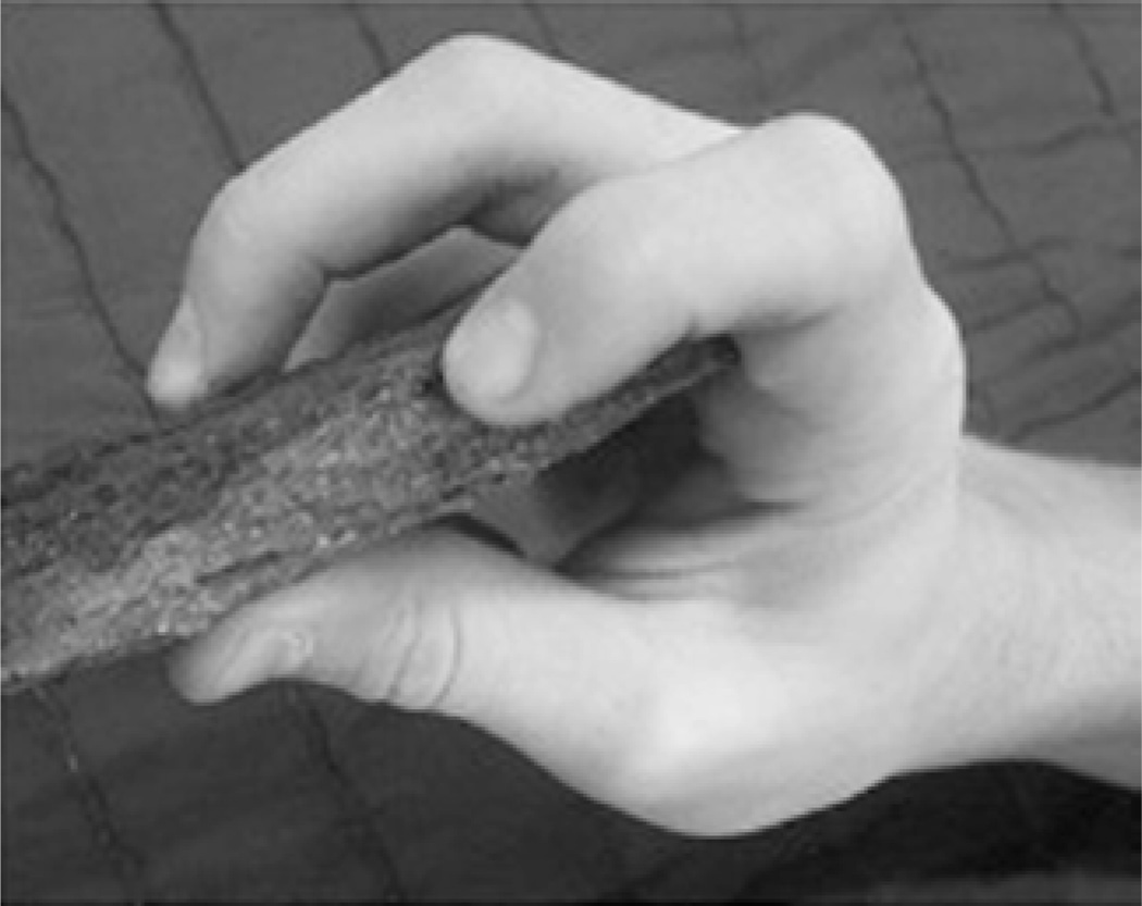
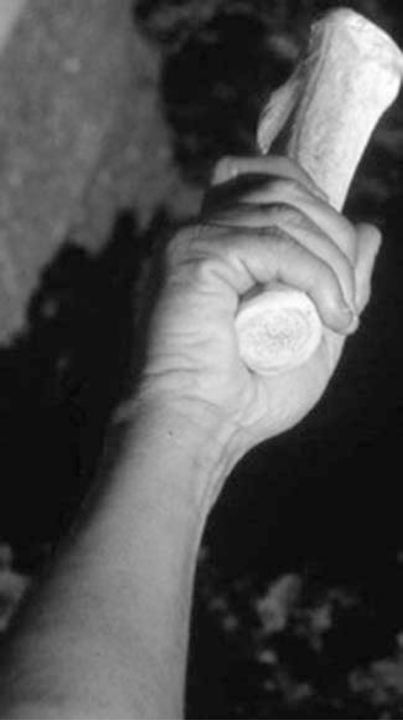
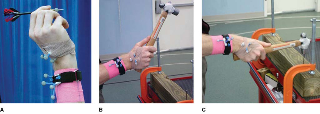
Unlike any other diarthrodial joint in the human body, the "wrist joint" is composed of numerous articulations between eight carpal bones, the distal radius, the distal ulna, and five metacarpal bones. The carpal bones articulate with each other as well as with the distal radius, distal ulna, and the metacarpal bases. Multiple theories explaining intercarpal motion have been proposed; however, controversy exists concerning the degree and direction of motion of the individual carpal bones within the two carpal rows during different planes of motion. Recent investigations have suggested that traditional explanations of carpal bone motion may not entirely account for carpal motion in all planes. Better understanding of the complexities of carpal motion through the use of advanced imaging techniques and simultaneous appreciation of human anatomic and functional evolution have led to the hypothesis that the "dart thrower's motion" of the wrist is uniquely human. Carpal kinematic research and current developments in both orthopaedic surgery and anthropology underscore the importance of the dart thrower's motion in human functional activities and the clinical implications of these concepts for orthopaedic surgery and rehabilitation.
Figures
Similar articles
-
Functional kinematics of the wrist.J Hand Surg Eur Vol. 2016 Jan;41(1):7-21. doi: 10.1177/1753193415616939. Epub 2015 Nov 14. J Hand Surg Eur Vol. 2016. PMID: 26568538 Review.
-
The effects of wrist distraction on carpal kinematics.J Hand Surg Am. 1999 Jan;24(1):113-20. doi: 10.1016/S0266-7681(99)90057-8. J Hand Surg Am. 1999. PMID: 10048525
-
A comparison of dart thrower's range of motion following radioscapholunate fusion, four-corner fusion and proximal row carpectomy.J Hand Surg Eur Vol. 2018 Sep;43(7):718-722. doi: 10.1177/1753193418783330. Epub 2018 Jun 27. J Hand Surg Eur Vol. 2018. PMID: 29950134
-
In-vivo three-dimensional carpal bone kinematics during flexion-extension and radio-ulnar deviation of the wrist: Dynamic motion versus step-wise static wrist positions.J Biomech. 2009 Dec 11;42(16):2664-71. doi: 10.1016/j.jbiomech.2009.08.016. Epub 2009 Sep 12. J Biomech. 2009. PMID: 19748626
-
International Federation of Societies for Surgery of the Hand 2013 Committee's report on wrist dart-throwing motion.J Hand Surg Am. 2014 Jul;39(7):1433-9. doi: 10.1016/j.jhsa.2014.02.035. J Hand Surg Am. 2014. PMID: 24888529 Review.
Cited by
-
Anatomy, Biomechanics, and Loads of the Wrist Joint.Life (Basel). 2022 Jan 27;12(2):188. doi: 10.3390/life12020188. Life (Basel). 2022. PMID: 35207475 Free PMC article. Review.
-
Carpal coalition: A review of current knowledge and report of a single institution's experience with asymptomatic intercarpal fusion.Hand (N Y). 2013 Jun;8(2):157-63. doi: 10.1007/s11552-013-9498-5. Hand (N Y). 2013. PMID: 24426912 Free PMC article.
-
Motion-plane dependency of the range of dart throw motion and the effects of tendon action due to finger extrinsic muscles during the motion.J Phys Ther Sci. 2018 Mar;30(3):355-360. doi: 10.1589/jpts.30.355. Epub 2018 Mar 2. J Phys Ther Sci. 2018. PMID: 29581651 Free PMC article.
-
Functional Dart-Throwing Motion: A Clinical Comparison of Four-Corner Fusion to Radioscapholunate Fusion Using Inertial Motion Capture.J Wrist Surg. 2020 Aug;9(4):321-327. doi: 10.1055/s-0040-1710500. Epub 2020 May 28. J Wrist Surg. 2020. PMID: 32760611 Free PMC article.
-
The forearm and hand musculature of semi-terrestrial rhesus macaques (Macaca mulatta) and arboreal gibbons (Fam. Hylobatidae). Part I. Description and comparison of the muscle configuration.J Anat. 2020 Oct;237(4):774-790. doi: 10.1111/joa.13222. Epub 2020 Jun 8. J Anat. 2020. PMID: 32511764 Free PMC article.
References
-
- Taleisnik J. The ligaments of the wrist. J Hand Surg Am. 1976;1:110–118. - PubMed
-
- Arkless R. Cineradiography in normal and abnormal wrists. Am J Roentgenol Radium Ther Nucl Med. 1966;96:837–844. - PubMed
-
- Werner FW, Short WH, Fortino MD, Palmer AK. The relative contribution of selected carpal bones to global wrist motion during simulated planar and out-of-plane wrist motion. J Hand Surg Am. 1997;22:708–713. - PubMed
Publication types
MeSH terms
Grants and funding
LinkOut - more resources
Full Text Sources
