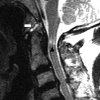
Revisiting anterior atlantoaxial subluxation with overlooked information on MR images
- PMID: 20044504
- PMCID: PMC7964179
- DOI: 10.3174/ajnr.A1941
Revisiting anterior atlantoaxial subluxation with overlooked information on MR images
Abstract
Background and purpose: The ADI is the imaging diagnostic clue to AAA subluxation of the cervical spine. Some MR imaging findings other than abnormal ADI relate to AAA subluxation. However, their relationship is not yet clarified. The present study elucidates the role of MR imaging by employing these previously overlooked findings.
Materials and methods: This study enrolled 40 patients with AAA subluxation and 20 non-AAA subluxation patients as controls. All MR imaging was performed with supine neutral positioning. The morphology of the dens, bilateral facet joints, and surrounding ligaments, as well as the alignment of the anterior atlantoaxial joint, the spinolaminar line, and the intramedullary signal intensity, were assessed. This investigation statistically analyzed the difference among these groups.
Results: Thirty-eight percent (15 of 40) of patients with AAA subluxation showed nAAA. There was no significant difference between the groups of AAA with normal and abnormal ADI except that more peridental pannus was seen in the latter group. More dens erosion (P = .022), tilting of anterior atlantoaxial joint (P = .022), peridental effusion (P < .001), lateral facet arthropathy (P < .001), abnormal spinolaminar line (P = .001), and focal myelopathy (P = .001) were observed in nAAA patients compared with the controls. The combination of peridental effusion, lateral facet arthropathy, abnormal intramedullary signals, and abnormal spinolaminar line showed a sensitivity of 100% and a specificity of 90% in diagnosing AAA subluxation.
Conclusions: MR imaging provides important biomechanical clues, other than ADI, that improve accuracy in diagnosing atlantoaxial instability.
Figures


Similar articles
-
Relationship between the morphology of the atlanto-occipital joint and the radiographic results in patients with atlanto-axial subluxation due to rheumatoid arthritis.Eur Spine J. 2008 Jun;17(6):826-30. doi: 10.1007/s00586-008-0659-0. Epub 2008 Apr 4. Eur Spine J. 2008. PMID: 18389289 Free PMC article.
-
Pre- and postoperative MR imaging of the craniocervical junction in rheumatoid arthritis.AJR Am J Roentgenol. 1989 Mar;152(3):561-6. doi: 10.2214/ajr.152.3.561. AJR Am J Roentgenol. 1989. PMID: 2783810
-
Clinical significance of articulating facet displacement of lateral atlantoaxial joint on 3D CT in diagnosing atlantoaxial subluxation.J Formos Med Assoc. 2007 Oct;106(10):840-6. doi: 10.1016/S0929-6646(08)60049-2. J Formos Med Assoc. 2007. PMID: 17964963
-
Imaging of Atlanto-Occipital and Atlantoaxial Traumatic Injuries: What the Radiologist Needs to Know.Radiographics. 2015 Nov-Dec;35(7):2121-34. doi: 10.1148/rg.2015150035. Radiographics. 2015. PMID: 26562241 Review.
-
Anteroposterior atlantoaxial subluxation in cervical spine osteoarthritis: case reports and review of the literature.J Rheumatol. 1999 Mar;26(3):687-91. J Rheumatol. 1999. PMID: 10090183 Review.
Cited by
-
Magnetic resonance imaging characteristics of atlanto-axial subluxation in 42 dogs: Analysis of joint cavity size, subluxation distance, and craniocervical junction anomalies.Open Vet J. 2023 Sep;13(9):1091-1098. doi: 10.5455/OVJ.2023.v13.i9.4. Epub 2023 Sep 30. Open Vet J. 2023. PMID: 37842109 Free PMC article.
-
Atlantoaxial Subluxation Related to Axial Spondylarthritis: A Case-Based Systematic Review.Mediterr J Rheumatol. 2024 Dec 31;35(4):563-572. doi: 10.31138/mjr.070624.asr. eCollection 2024 Dec. Mediterr J Rheumatol. 2024. PMID: 39886282 Free PMC article. Review.
-
Comparison of the diagnostic performance of the Swischuk line method and the anterior atlantodental interval method in atlantodental subluxation.BMC Med Imaging. 2024 Jan 2;24(1):8. doi: 10.1186/s12880-023-01187-z. BMC Med Imaging. 2024. PMID: 38166926 Free PMC article.
-
Grisel's Syndrome After COVID-19 in a Pediatric Patient: A Case Report.Cureus. 2024 Jun 9;16(6):e62028. doi: 10.7759/cureus.62028. eCollection 2024 Jun. Cureus. 2024. PMID: 38989331 Free PMC article.
-
Radiological Evaluation of Cervical Spine Involvement in Rheumatoid Arthritis: A Cross-Sectional Retrospective Study.J Clin Med. 2021 Oct 5;10(19):4587. doi: 10.3390/jcm10194587. J Clin Med. 2021. PMID: 34640605 Free PMC article.
References
-
- Zikou AK, Alamanos Y, Argyropoulou MI, et al. . Radiological cervical spine involvement in patients with rheumatoid arthritis: a cross sectional study. J Rheumatol 2005;32:801–06 - PubMed
-
- Naranjo A, Carmona L, Gavrila D, et al. . Prevalence and associated factors of anterior atlantoaxial luxation in a nation-wide sample of rheumatoid arthritis patients. Clin Exp Rheumatol 2004;22:427–32 - PubMed
-
- Kauppi M, Hakala M. Prevalence of cervical spine subluxations and dislocations in a community-based rheumatoid arthritis population. Scand J Rheumatol 1994;23:133–36 - PubMed
-
- Brattstrom H, Granholm L. Atlanto-axial fusion in rheumatoid arthritis: a new method of fixation with wire and bone cement. Acta Orthop Scand 1976;47:619–28 - PubMed
Publication types
MeSH terms
LinkOut - more resources
Full Text Sources
Medical
