

MR imaging of adenomas of the nonpigmented ciliary epithelium of the eye
- PMID: 20044507
- PMCID: PMC7964188
- DOI: 10.3174/ajnr.A1947
MR imaging of adenomas of the nonpigmented ciliary epithelium of the eye
Abstract
Background and purpose: ANPCEs are rare benign tumors of the eye arising from the NPCE in adults, which may be clinically mistaken for melanoma. This study was undertaken to delineate clinical and MR imaging features of these tumors.
Materials and methods: Clinical presentation and MR imaging findings of 8 patients (6 women and 2 men; median age, 51 years) with pathologically confirmed ANPCEs were retrospectively reviewed. Location, size, shape, margin, signal intensity, and gadolinium-enhancement characteristics of all tumors were evaluated. Signal intensity and degree of enhancement were graded in comparison with the ipsilateral lacrimal gland.
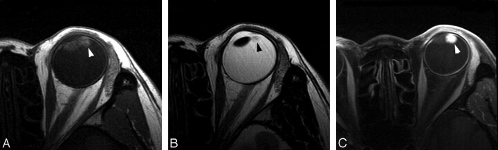
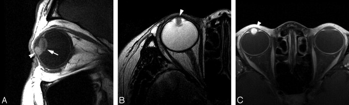
Results: MR imaging revealed a circumscribed enhancing mass within the ciliary body of the eye in all 8 patients. The mass was ovoid in 6 patients and spheric in 2. Gadolinium enhancement was marked in 4 lesions and moderate in the other 4. Both T1 and T2 relaxation times were qualitatively identical to those in the lacrimal gland in 2 tumors. In the remaining 6 tumors, the T1 was identical to and the T2 longer than that in the lacrimal gland.
Conclusions: ANPCE should be included in the differential diagnosis of a spheric or ovoid enhancing ciliary body mass with T1 similar to that in the lacrimal gland and T2 equal to or longer than that in the lacrimal gland.
Figures
Similar articles
-
Acquired adenoma of the nonpigmented ciliary epithelium: analysis of five cases.Graefes Arch Clin Exp Ophthalmol. 2015 Apr;253(4):637-44. doi: 10.1007/s00417-014-2928-4. Epub 2015 Jan 22. Graefes Arch Clin Exp Ophthalmol. 2015. PMID: 25605545
-
Acquired neoplasms of the nonpigmented ciliary epithelium (adenoma and adenocarcinoma).Ophthalmology. 1996 Dec;103(12):2007-16. doi: 10.1016/s0161-6420(96)30393-x. Ophthalmology. 1996. PMID: 9003334
-
Clinical and histopathological features of adenomas of the ciliary pigment epithelium.Acta Ophthalmol. 2016 Nov;94(7):e637-e643. doi: 10.1111/aos.13029. Epub 2016 Apr 29. Acta Ophthalmol. 2016. PMID: 27130243
-
Adenoma of nonpigmented epithelium in ciliary body: literature review and case report.J Zhejiang Univ Sci B. 2007 Sep;8(9):612-5. doi: 10.1631/jzus.2007.B0612. J Zhejiang Univ Sci B. 2007. PMID: 17726740 Free PMC article. Review.
-
TUMORS OF THE NONPIGMENTED EPITHELIUM OF THE CILIARY BODY: The Lorenz E. Zimmerman Tribute Lecture.Retina. 2015 May;35(5):957-65. doi: 10.1097/IAE.0000000000000445. Retina. 2015. PMID: 25545484 Review.
Cited by
-
Acquired adenoma of the nonpigmented ciliary epithelium: analysis of five cases.Graefes Arch Clin Exp Ophthalmol. 2015 Apr;253(4):637-44. doi: 10.1007/s00417-014-2928-4. Epub 2015 Jan 22. Graefes Arch Clin Exp Ophthalmol. 2015. PMID: 25605545
-
Rare Histological Type of Adenoma of the Nonpigmented Ciliary Epithelium.Case Rep Ophthalmol. 2019 Feb 20;10(1):75-80. doi: 10.1159/000497033. eCollection 2019 Jan-Apr. Case Rep Ophthalmol. 2019. PMID: 31097948 Free PMC article.
References
-
- Appolloni R, Modesti M, Pecorella I, et al. . Uncommon cause of juvenile cataract: adenoma of the nonpigmented ciliary epithelium. J Cataract Refract Surg 2008;34:1997–2001 - PubMed
-
- Elizalde J, Ubia S, Barraquer RI. Adenoma of the nonpigmented ciliary epithelium. Eur J Ophthalmol 2006;16:630–33 - PubMed
-
- Murphy MF, Johnston PB, Lyness RW. Adenoma of the non-pigmented epithelium of the ciliary body. Eye 1997;11:419–20 - PubMed
-
- Cursiefen C, Schlötzer-Schrehardt U, Holbach LM, et al. . Adenoma of the nonpigmented ciliary epithelium mimicking a malignant melanoma of the iris. Arch Ophthalmol 1999;117:113–16 - PubMed
Publication types
MeSH terms
LinkOut - more resources
Full Text Sources
Medical