

Retinoid-induced histone deacetylation inhibits telomerase activity in estrogen receptor-negative breast cancer cells
- PMID: 20044602
- PMCID: PMC2888005
Retinoid-induced histone deacetylation inhibits telomerase activity in estrogen receptor-negative breast cancer cells
Abstract
Background: Multiple mechanisms regulate cancer-associated telomerase activity at the level of human telomerase reverse transcriptase (hTERT) transcription which may serve as novel targets for anticancer approaches.
Materials and methods: The effects of prolonged all-trans retinoic acid (ATRA) exposure on hTERT regulation in estrogen receptor-negative SK-BR-3 breast cancer cells were examined.
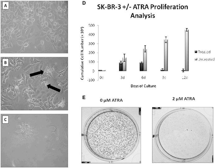
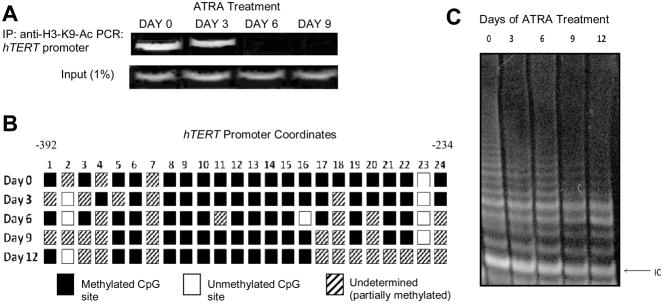
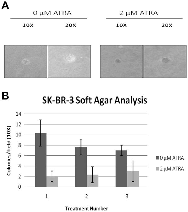
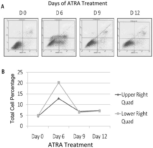
Results: ATRA had a profound effect on the morphology and proliferation rate of the SK-BR-3 cells. ATRA also hindered the ability of these cancer cells to grow independently, rendering them more like normal somatic cells. The effect of ATRA on the decrease of telomerase activity was found to be associated with a rapid decrease in histone H3-lysine 9 acetylation (H3-K9-Ac) of the hTERT promoter. Extended-exposure to ATRA in these cells also caused the initiation of a putative compensatory mechanism, counteracting the induced surge in apoptosis.
Conclusion: A rapid decrease of H3-K9 acetylation at the hTERT promoter could be an important mechanism by which ATRA shuts down telomerase activity and mediates its antitumor effects in estrogen receptor-negative breast cancer cells.
Figures
Similar articles
-
All-trans-retinoic acid modulates glycolysis via H19 and telomerase: the role of mir-let-7a in estrogen receptor-positive breast cancer cells.BMC Cancer. 2024 May 21;24(1):615. doi: 10.1186/s12885-024-12379-3. BMC Cancer. 2024. PMID: 38773429 Free PMC article.
-
Sulforaphane causes epigenetic repression of hTERT expression in human breast cancer cell lines.PLoS One. 2010 Jul 6;5(7):e11457. doi: 10.1371/journal.pone.0011457. PLoS One. 2010. PMID: 20625516 Free PMC article.
-
Lysine-specific demethylase 1 (LSD1) Is required for the transcriptional repression of the telomerase reverse transcriptase (hTERT) gene.PLoS One. 2008 Jan 16;3(1):e1446. doi: 10.1371/journal.pone.0001446. PLoS One. 2008. PMID: 18197256 Free PMC article.
-
Sulforaphane modulates telomerase activity via epigenetic regulation in prostate cancer cell lines.Biochem Cell Biol. 2016 Feb;94(1):71-81. doi: 10.1139/bcb-2015-0038. Epub 2015 Sep 9. Biochem Cell Biol. 2016. PMID: 26458818
-
DNA Methylation Profiling of hTERT Gene Alongside with the Telomere Performance in Gastric Adenocarcinoma.J Gastrointest Cancer. 2020 Sep;51(3):788-799. doi: 10.1007/s12029-020-00427-7. J Gastrointest Cancer. 2020. PMID: 32617831 Review.
Cited by
-
Autophagy inhibition synergistically enhances anticancer efficacy of RAMBA, VN/12-1 in SKBR-3 cells, and tumor xenografts.Mol Cancer Ther. 2012 Apr;11(4):898-908. doi: 10.1158/1535-7163.MCT-11-0860. Epub 2012 Feb 14. Mol Cancer Ther. 2012. PMID: 22334589 Free PMC article.
-
Combining old and new concepts in targeting telomerase for cancer therapy: transient, immediate, complete and combinatory attack (TICCA).Cancer Cell Int. 2023 Sep 7;23(1):197. doi: 10.1186/s12935-023-03041-2. Cancer Cell Int. 2023. PMID: 37679807 Free PMC article. Review.
-
Involvement of epigenetic modification of TERT promoter in response to all-trans retinoic acid in ovarian cancer cell lines.J Ovarian Res. 2019 Jul 10;12(1):62. doi: 10.1186/s13048-019-0536-y. J Ovarian Res. 2019. PMID: 31291979 Free PMC article.
-
Telomerase Regulation from Beginning to the End.Genes (Basel). 2016 Sep 14;7(9):64. doi: 10.3390/genes7090064. Genes (Basel). 2016. PMID: 27649246 Free PMC article. Review.
-
Telomerase as a useful target in cancer fighting-the breast cancer case.Tumour Biol. 2013 Jun;34(3):1371-80. doi: 10.1007/s13277-013-0757-4. Epub 2013 Apr 5. Tumour Biol. 2013. PMID: 23558965 Free PMC article. Review.
References
-
- Yang L, Tin-U C, Wu L, Brown P. Role of retinoid receptors in the prevention and treatment of breast cancer. J Mammary Gland Biol Neoplasia. 1999;4:377–388. - PubMed
-
- Roman S, Clarke C, Hall R, Alexander I, Sutherland R. Expression and regulation of retinoic acid receptors in human breast cancer cells. Cancer Res. 1992;52:2236–2242. - PubMed
-
- van der Burg B, van der Leede BM, Kwakkenbos-Isbrucker L, Salverda S, de Laat SW, van der Saag PT. Retinoic acid resistance of estradiol-independent breast cancer cells coincides with diminished retinoic acid receptor function. Mol Cell Endocrinol. 1993;91:149–157. - PubMed
-
- Krupitza G, Hulla W, Harant H, Dittrich E, Kallay E, Huber H, Grunt T, Dittrich C. Retinoic acid induced death of ovarian carcinoma cells correlates with c-myc stimulation. Int J Cancer. 1995;61:649–657. - PubMed
Publication types
MeSH terms
Substances
Grants and funding
LinkOut - more resources
Full Text Sources
Medical