. 2010 Jul;49(1):70-8.
doi: 10.1016/j.yjmcc.2009.12.013. Epub 2010 Jan 4.

Tumor necrosis factor-alpha produced in cardiomyocytes mediates a predominant myocardial inflammatory response to stretch in early volume overload

Affiliations

Tumor necrosis factor-alpha produced in cardiomyocytes mediates a predominant myocardial inflammatory response to stretch in early volume overload

Yuanwen Chen et al. J Mol Cell Cardiol. 2010 Jul.

Abstract

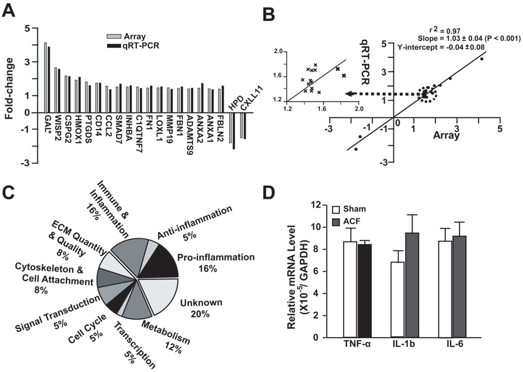
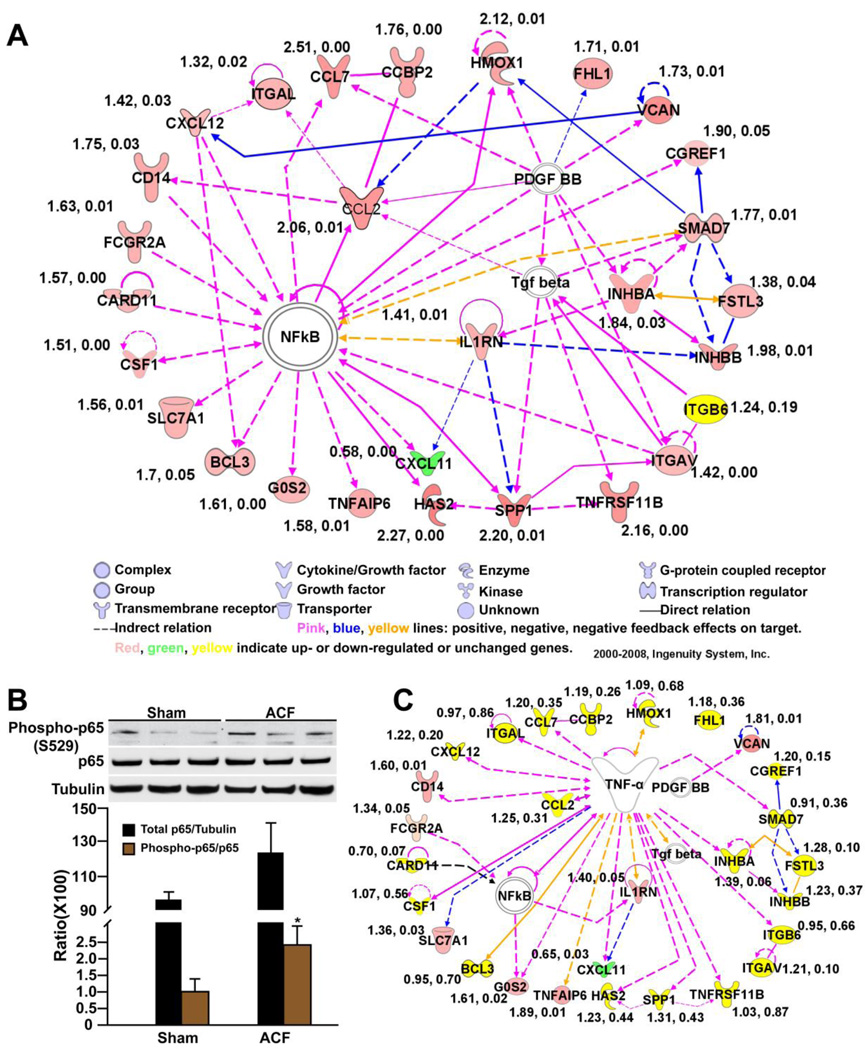
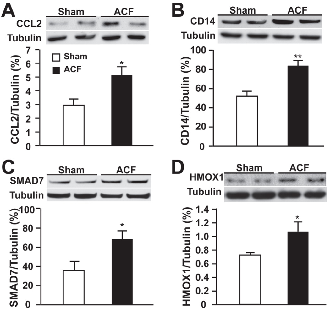
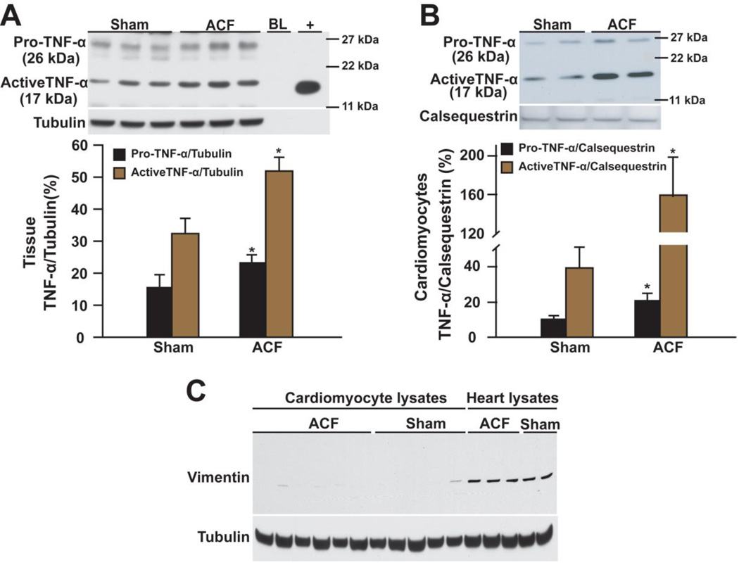
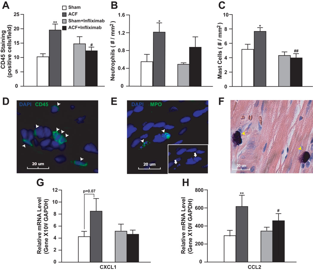
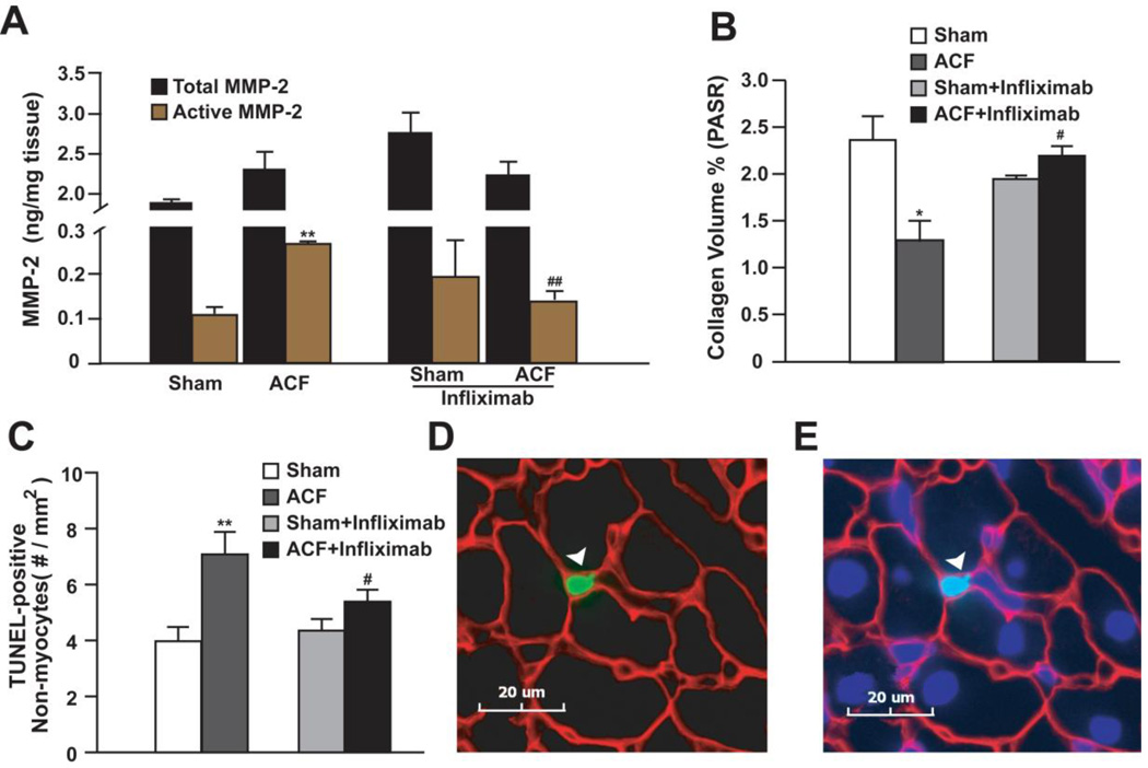
Acute stretch caused by volume overload (VO) of aorto-caval fistula (ACF) induces a variety of myocardial responses including mast cell accumulation, matrix metalloproteinase (MMP) activation, and collagen degradation, all of which are critical in dictating long-term left ventricle (LV) outcome to VO. Meanwhile, these responses can be part of myocardial inflammation dictated by tumor necrosis factor-alpha (TNF-alpha), which is elevated after acute ACF. However, it is unknown whether TNF-alpha mediates a major myocardial inflammatory response to stretch in early VO. In 24-h ACF and sham rats, microarray gene expression profiling and subsequent Ingenuity Pathway Analysis identified a predominant inflammatory response and a gene network of biologically interactive genes strongly linked to TNF-alpha. Western blot demonstrated increased local production of TNF-alpha in the LV (1.71- and 1.66-fold in pro- and active-TNF-alpha over control, respectively, P<0.05) and cardiomyocytes (2- and 4-fold in pro- and active-TNF-alpha over control, respectively, P<0.05). TNF-alpha neutralization with infliximab (5.5 mg/kg) attenuated the myocardial inflammatory response to acute VO, as indicated by inhibition of inflammatory gene upregulation, myocardial infiltration (total CD45+ cells, mast cells, and neutrophils), MMP-2 activation, collagen degradation, and cardiac cell apoptosis, without improving LV remodeling and function. These results indicate that TNF-alpha produced by cardiomyocytes mediates a predominant inflammatory response to stretch in the early VO in the ACF rat, suggesting an important role of TNF-alpha in initiating pathophysiological response of myocardium to VO.


Figures

Figure 1
Figure 1. Microarray analysis and validation by qRT-PCR of 24 hour ACF
(A) Comparison of gene fold-change by microarray and qRT-PCR in 10% of the total altered genes identified by microarray. *Refers to supplemental Table 1 for gene annotation. (B) Correlation between expression changes by microarray vs. qRT-PCR with best-fit linear regression line. Inset - Genes with a 1.4-2-fold change plotted for better visibility. (C) Functional classification of genes (162) differentially expressed in ACF vs. Sham. (D) Relative expression of TNF-α, IL-1b (interleukin 1β) and IL-6 (interleukin 6) mRNA by qRT-PCR in ACF and sham.
Figure 2
Figure 2. IPA analysis of differentially expressed genes in 24 hour ACF (see Table 2)
(A) IPA network of interactive genes involved in inflammation and immune response (P < 10−56). Connecting line(s) represent known biological relation between genes. Fold-changes and P-value derived from qRT-PCR are listed beside each gene. (B) Western blot of phospho- and total NF-κB p65 in ACF vs. sham. Tubulin loading control. *P < 0.05 vs. sham. (C) IPA analysis of the interaction between TNF-α and the identified gene network in (A). Effect of TNF-α neutralization on each gene was determined by qRT-PCR (fold-change and P-value listed beside each gene).
Figure 3
Figure 3. Representative western blot of TNF-α related inflammation proteins in IPA network
Expression of (A) CCL2 (chemokine (c-c) motif ligand 2), (B) CD14, (C) SMAD7 (SMAD family member 7) and (D) HMOX1 (heme oxygenase 1) in sham and ACF at 24 hours. Densitometric analysis after normalization to tubulin is presented below. *P < 0.05, **P < 0.01 vs. sham.
Figure 4
Figure 4. Western blot of TNF-α in LV tissue and isolated cardiomyocytes of 24 hour ACF
Pro- and active TNF-α in (A) heart lysates and (B) cardiomyocytes from ACF vs. shams. “BL” loading dye. “+” recombinant active rat TNF-α. Densitometric analysis is presented below. *P < 0.05, **P < 0.01 vs. sham. (C) Western blot of vimentin in cardiomyocytes and heart lysates (positive for vimentin) to ensure the purity of cardiomyocytes. Tubulin or calsequestrin was used as loading control.
Figure 5
Figure 5. Effect of TNF-α neutralization (Infliximamb) on myocardial inflammatory response to 24 hour ACF
Number of (A) CD45+ cells, (B) neutrophils and (C) mast cells in ACF and shams with or without infliximab was quantified. Representative images of (D) CD45+ cells (green), (E) neutrophils with myeloperoxidase (MPO) staining (green), and (F) mast cells with Giemsa staining (purple) were shown below. Nuclei were stained blue with DAPI. Arrowheads indicate positively stained cells. Arrows in (E) demonstrate multilobular nuclei of neutrophils. (G) and (H) demonstrate relative CXCL1 and CCL2 mRNA levels quantified by qRT-PCR in ACF and shams with or without infliximab. Expression level was normalized to GAPDH. *P<0.05, **P< 0.01 vs. sham; #P<0.05, ##P<0.01 vs. ACF.
Figure 6
Figure 6. Effect of TNF-α neutralization on MMP-2, collagen and apoptosis in 24 hour ACF
(A) LV total and active MMP-2 and (B) interstitial collagen in ACF and sham rats with or without infliximab was quantified. (C) Quantification of TUNEL-positive non-myocyte nuclei in ACF and sham rats with or without infliximab. (D) Representative image of a TUNEL-positive non-cardiomyocyte nucleus (green - arrowhead) (E) also nuclear stained with DAPI (blue). Counter stain is laminin (red). *P<0.05, **P< 0.01 vs. sham; #P<0.05, ##P<0.01 vs. ACF.

References

    1. Brower GL, Janicki JS. Pharmacologic inhibition of mast cell degranulation prevents left ventricular remodeling induced by chronic volume overload in rats. J Card Fail. 2005;11:548–556. - PubMed
    1. Ryan TD, Rothstein EC, Aban I, Tallaj JA, Husain A, Lucchesi PA, et al. Left ventricular eccentric remodeling and matrix loss are mediated by bradykinin and precede cardiomyocyte elongation in rats with volume overload. J Am Coll Cardiol. 2007;49:811–821. - PubMed
    1. Levick SP, Gardner JD, Holland M, Hauer-Jensen M, Janicki JS, Brower GL. Protection from adverse myocardial remodeling secondary to chronic volume overload in mast cell deficient rats. J Mol Cell Cardiol. 2008;45:56–61. - PMC - PubMed
    1. Brower GL, Chancey AL, Thanigaraj S, Matsubara BB, Janicki JS. Cause and effect relationship between myocardial mast cell number and matrix metalloproteinase activity. Am J Physiol Heart Circ Physiol. 2002;283:H518–H525. - PubMed
    1. Kubota T, McTiernan CF, Frye CS, Demetris AJ, Feldman AM. Cardiac-specific overexpression of tumor necrosis factor-alpha causes lethal myocarditis in transgenic mice. J Card Fail. 1997;3:117–124. - PubMed

Publication types

MeSH terms

Substances