

RSK in tumorigenesis: connections to steroid signaling
- PMID: 20045011
- PMCID: PMC2823981
- DOI: 10.1016/j.steroids.2009.12.010
RSK in tumorigenesis: connections to steroid signaling
Abstract
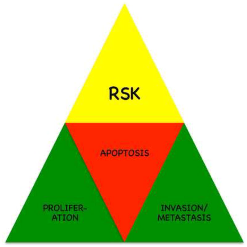
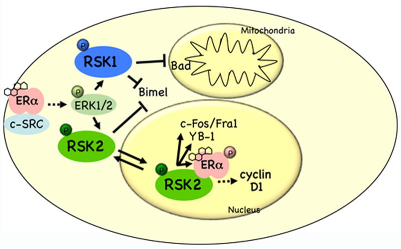
The Ser/Thr kinase family, RSK, has been implicated in numerous types of hormone-dependent and -independent cancers. However, there has been little consideration of RSKs as downstream mediators of steroid hormone non-genomic effects or of their ability to facilitate steroid receptor-mediated gene expression. Steroid hormone signaling can directly stimulate the MEK/ERK/RSK pathway to regulate cellular proliferation and survival in transformed cells. To date, multiple mechanisms of RSK and steroid hormone receptor-mediated proliferation/survival have been elucidated. For example, RSK enhances proliferation of breast and prostate cancer cells via its ability to control the levels of the estrogen receptor co-activator, cyclin D1. While in lung and other tumors RSK may control apoptosis via estrogen-mediated regulation of mitochondrial integrity. Thus the RSKs could be important anti-cancer therapeutic targets in many different transformed tissues. The recent discovery of RSK-specific inhibitors will advance our current understanding of RSK in transformation and drive these studies into animal and clinical models. In this review we explore the mechanisms associated with RSK in tumorigenesis and their relationship to steroid hormone signaling.
Copyright 2009 Elsevier Inc. All rights reserved.
Figures
Similar articles
-
Defining the role of the RSK isoforms in cancer.Semin Cancer Biol. 2018 Feb;48:53-61. doi: 10.1016/j.semcancer.2017.04.016. Epub 2017 May 2. Semin Cancer Biol. 2018. PMID: 28476656 Review.
-
The p90 ribosomal S6 kinase (RSK) inhibitor BI-D1870 prevents gamma irradiation-induced apoptosis and mediates senescence via RSK- and p53-independent accumulation of p21WAF1/CIP1.Cell Death Dis. 2013 Oct 17;4(10):e859. doi: 10.1038/cddis.2013.386. Cell Death Dis. 2013. PMID: 24136223 Free PMC article.
-
RSK-mediated down-regulation of PDCD4 is required for proliferation, survival, and migration in a model of triple-negative breast cancer.Oncotarget. 2016 May 10;7(19):27567-83. doi: 10.18632/oncotarget.8375. Oncotarget. 2016. PMID: 27028868 Free PMC article.
-
RSK activation via ERK modulates human colon cancer cells response to PTHrP.J Mol Endocrinol. 2017 Jul;59(1):13-27. doi: 10.1530/JME-16-0216. Epub 2017 Apr 6. J Mol Endocrinol. 2017. PMID: 28385776
-
Role and regulation of 90 kDa ribosomal S6 kinase (RSK) in signal transduction.Mol Cell Endocrinol. 1999 May 25;151(1-2):65-77. doi: 10.1016/s0303-7207(99)00061-1. Mol Cell Endocrinol. 1999. PMID: 10411321 Review.
Cited by
-
An oral first-in-class small molecule RSK inhibitor suppresses AR variants and tumor growth in prostate cancer.Cancer Sci. 2022 May;113(5):1731-1738. doi: 10.1111/cas.15280. Epub 2022 Apr 1. Cancer Sci. 2022. PMID: 35118769 Free PMC article.
-
Myricetin exerts anti-proliferative, anti-invasive, and pro-apoptotic effects on esophageal carcinoma EC9706 and KYSE30 cells via RSK2.Tumour Biol. 2014 Dec;35(12):12583-92. doi: 10.1007/s13277-014-2579-4. Epub 2014 Sep 6. Tumour Biol. 2014. PMID: 25192723
-
The unusual mechanism of inhibition of the p90 ribosomal S6 kinase (RSK) by flavonol rhamnosides.Biochim Biophys Acta. 2013 Jul;1834(7):1285-91. doi: 10.1016/j.bbapap.2013.03.018. Epub 2013 Mar 27. Biochim Biophys Acta. 2013. PMID: 23541530 Free PMC article. Review.
-
Phosphorylation of p90RSK is associated with increased response to neoadjuvant chemotherapy in ER-positive breast cancer.BMC Cancer. 2012 Dec 10;12:585. doi: 10.1186/1471-2407-12-585. BMC Cancer. 2012. PMID: 23216670 Free PMC article.
-
Amphiregulin Is a Critical Downstream Effector of Estrogen Signaling in ERα-Positive Breast Cancer.Cancer Res. 2015 Nov 15;75(22):4830-8. doi: 10.1158/0008-5472.CAN-15-0709. Epub 2015 Nov 2. Cancer Res. 2015. PMID: 26527289 Free PMC article.
References
-
- Shupnik MA. Crosstalk between steroid receptors and the c-Src-receptor tyro ine kinase pathways: implications for cell proliferation. Oncogene. 2004;23:7979–7989. - PubMed
-
- Cheskis BJ, Greger J, Cooch N, McNally C, McLarney S, Lam HS, Rutledge S, Mekonnen B, Hauze D, Nagpal S, et al. MNAR plays an important role in ERa activation of Src/MAPK and PI3K/Akt signaling pathways. Steroids. 2008;73:901–905. - PubMed
-
- Sebolt-Leopold JS, Herrera R. Targeting the mitogen-activated protein kinase cascade to treat cancer. Nat Rev Cancer. 2004;4:937–947. - PubMed
-
- Dougherty MK, Muller J, Ritt DA, Zhou M, Zhou XZ, Copeland TD, Conrads TP, Veenstra TD, Lu KP, Morrison DK. Regulation of Raf-1 by direct feedback phosphorylation. Mol Cell. 2005;17:215–224. - PubMed
Publication types
MeSH terms
Substances
Grants and funding
LinkOut - more resources
Full Text Sources
Research Materials
Miscellaneous
