. 2010 May;171(1):40-4.
doi: 10.1016/j.molbiopara.2009.12.009. Epub 2010 Jan 4.

A dispensable Plasmodium locus for stable transgene expression

Affiliations

A dispensable Plasmodium locus for stable transgene expression

Vanessa Y Jacobs-Lorena et al. Mol Biochem Parasitol. 2010 May.

Abstract

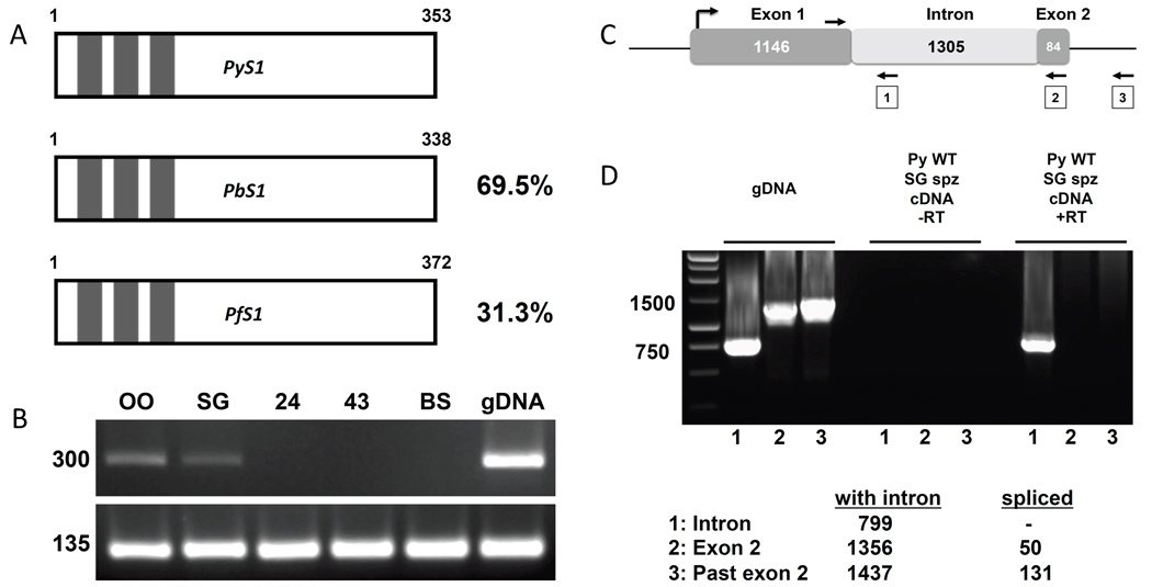
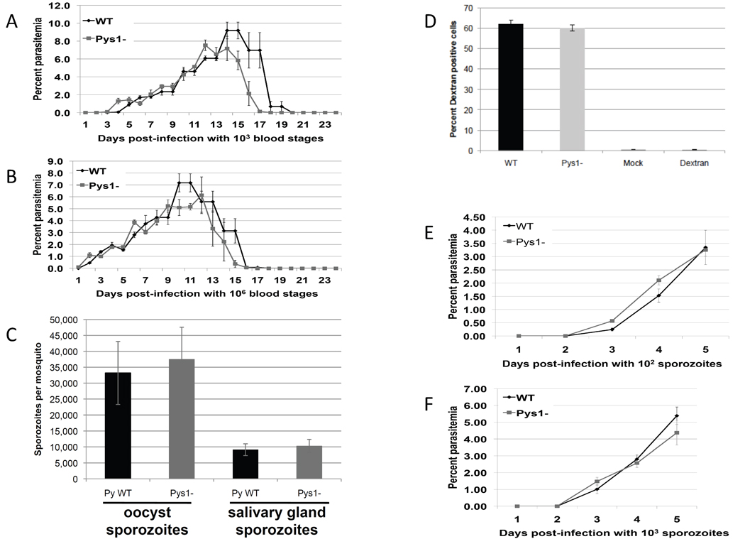
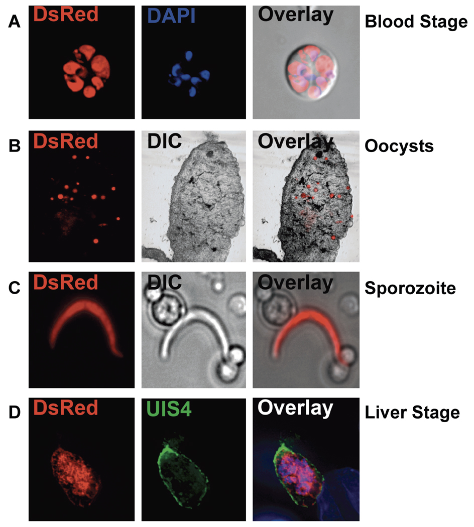
The ribosomal small subunit locus has been used for transgene expression in the rodent malaria parasites, Plasmodium berghei and Plasmodium yoelii, but this strategy utilizes single crossover integration and is thus prone to reversion by plasmid excision. Targeting of the ribosomal subunit locus may also have a negative effect on oocyst development in the mosquito. In P. berghei, the p230 paralog locus has been used for transgene expression. Here, we show that the P. yoelii S1 locus (sporozoite expressed gene 1) (PY05712) is dispensable and can be used for stable transgene expression throughout the parasite life cycle. P. yoelii s1(-) parasites show no defect in blood stage replication, oocyst formation, sporozoite production, or liver stage development when compared to P. yoelii wildtype parasites. Further, we show that a fluorescent transgene can be stably expressed from this site. This demonstrates that the S1 locus can be utilized for stable expression of heterologous genes in rodent malaria parasites.


Figures

Figure 1
Figure 1
P. yoelii S1 gene structure (A) A schematic representation of S1 from P. yoelii, P. berghei, and P. falciparum. Length of the predicted protein is noted in amino acids, percentages indicate amino acid identity as compared to P. yoelii, and C3H1 zinc finger domains are represented by boxes. (B) PCR analysis showing transcription during oocyst sporozoites (OO), salivary gland sporozoites (SG), and no transcription during liver stages at 24 and 43 hrs post-infection (24, 43) and mixed blood stages (BS). Amplification of Py18S was used as a positive control. (C) Schematic representation of the PyS1 locus as annotated in PlasmoDB. The arrows denote the primers used to test the intron/exon structure of PyS1. (D) PCR analysis showing that PyS1 has a single uninterrupted ORF. Lanes labeled “1” are products from the reverse primer in the putative intron, lanes labeled “2” are products from the reverse primer in the putative second exon, lanes labeled “3” are products from the reverse primer beyond the putative second exon. Expected amplicon sizes are shown in the legend.
Figure 2
Figure 2
Phenotypic analysis of Pys1 parasites. Either 1000 (A) or one million (B) Pys1 or WT blood stage parasites were injected into four BALB/c mice. Blood stage parasitemia was followed every day for 24 days and expressed as an average percentage. (C) Pys1 and WT infected mosquitoes were evaluated for numbers of midgut and salivary gland sporozoites at day 10 and 14, respectively. (D) P. yoelii wildtype parasites (WT), Pys1 parasites, or uninfected mosquito salivary gland material (Mock) or media supplemented with dextran only (Dextran) were incubated with hepatoma cells for one hour. Cells were then trypsinized and counted via fluorescence-activated cell sorting to determine the percentage of cells that were dextran positive. (E) 100 or (F) 1,000 PyS1 or WT sporozoites were injected IV into four BALB/c mice. Blood stage parasitemia was followed every day for five days and expressed as an average percentage.
Figure 3
Figure 3
Fluorescence microscopy of Pys1 parasites. Expression of DsRed in Pys1 parasites was evident in (A) live blood stages whose nuclei were visualized with 4',6-diamidino-2-phenylindole (DAPI) (Blue), (B) mosquito midgut oocysts in freshly dissected midguts, (C) the salivary gland sporozoite released from dissected mosquito salivary glands by grinding, and (D) liver stages in hepatoma cells fixed and visualized with an anti-UIS4 antibody and DAPI. Fluorescent and differential interference contrast (DIC) images were captured and merged (Overlay).

References

    1. Malaria Fact Sheet no. 94 on World Wide Web URL: http://www.who.int/mediacentre/factsheets/fs094/en/index.html.
    1. Vaughan AM, Aly AS, Kappe SH. Malaria parasite pre-erythrocytic stage infection: gliding and hiding. Cell Host Microbe. 2008;4:209–218. - PMC - PubMed
    1. Sinnis P, Coppi A. A long and winding road: the Plasmodium sporozoite's journey in the mammalian host. Parasitol Int. 2007;56:171–178. - PMC - PubMed
    1. Mikolajczak SA, Kappe SH. A clash to conquer: the malaria parasite liver infection. Mol Microbiol. 2006;62:1499–1506. - PubMed
    1. Baton LA, Ranford-Cartwright LC. Spreading the seeds of million-murdering death: metamorphoses of malaria in the mosquito. Trends Parasitol. 2005;21:573–580. - PubMed

Publication types

MeSH terms

Substances

LinkOut - more resources