. 2010 Mar;37(3):764-71.
doi: 10.1016/j.nbd.2009.12.020. Epub 2010 Jan 4.

Modulation of astrocyte glutamate transporters decreases seizures in a mouse model of Tuberous Sclerosis Complex

Affiliations

Modulation of astrocyte glutamate transporters decreases seizures in a mouse model of Tuberous Sclerosis Complex

Ling-Hui Zeng et al. Neurobiol Dis. 2010 Mar.

Abstract

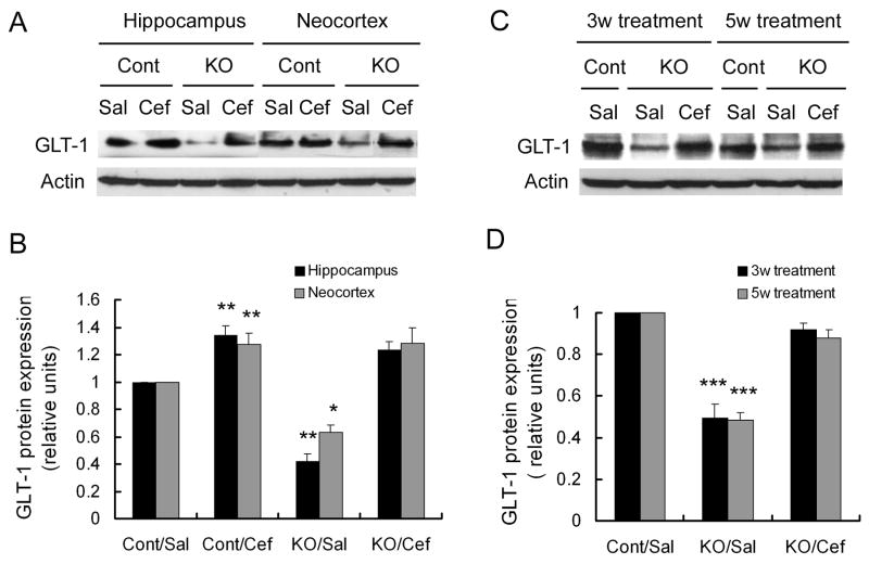
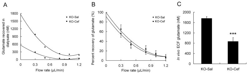
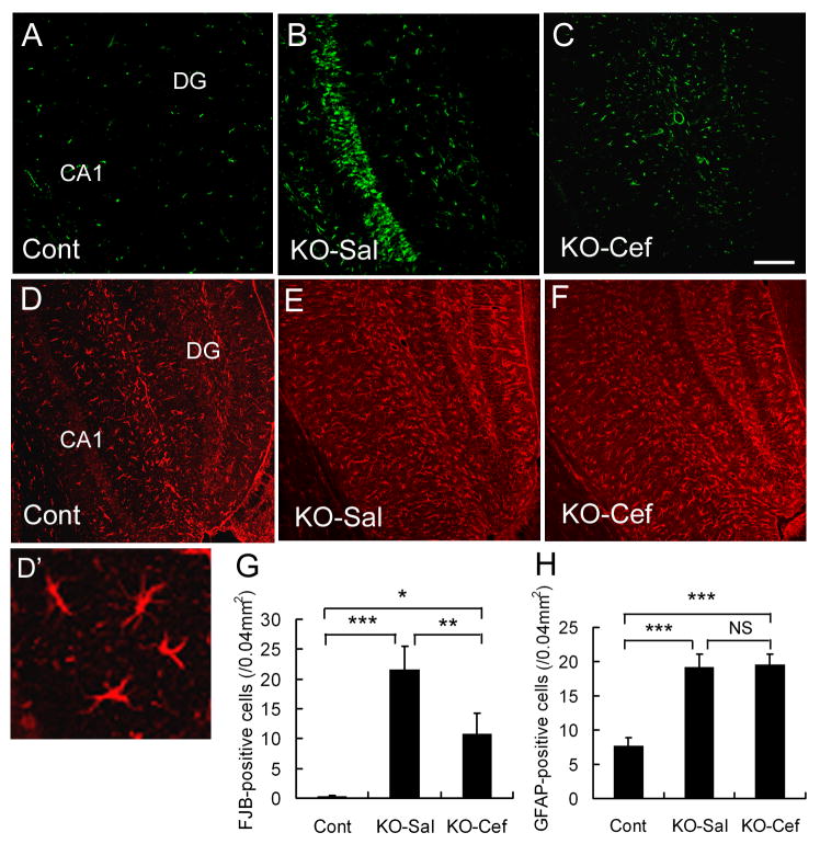
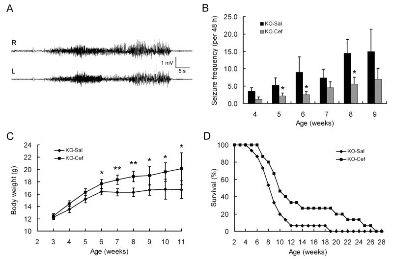
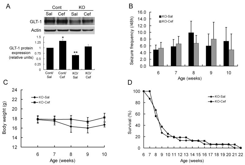
Astrocyte dysfunction may contribute to epileptogenesis and other neurological deficits in Tuberous Sclerosis Complex (TSC). In particular, decreased expression and function of astrocyte glutamate transporters have been implicated in causing elevated extracellular glutamate levels, neuronal death, and epilepsy in a mouse model of TSC (Tsc1(GFAP)CKO mice), involving inactivation of the Tsc1 gene primarily in astrocytes. Here, we tested whether pharmacological induction of astrocyte glutamate transporter expression can prevent the neurological phenotype of Tsc1(GFAP)CKO mice. Early treatment with ceftriaxone prior to the onset of epilepsy increased expression of astrocyte glutamate transporters, decreased extracellular glutamate levels, neuronal death, and seizure frequency, and improved survival in Tsc1(GFAP)CKO mice. In contrast, late treatment with ceftriaxone after onset of epilepsy increased glutamate transporter expression, but had no effect on seizures. These results indicate that astrocyte glutamate transporters contribute to epileptogenesis in Tsc1(GFAP)CKO mice and suggest novel therapeutic strategies for epilepsy in TSC directed at astrocytes.


Figures

Figure 1
Figure 1
Ceftriaxone increases astrocyte GLT-1 expression. (A) Three week old Tsc1GFAPCKO (KO) and control (Cont) mice received daily injections of ceftriaxone (Cef) or saline (Sal) for one week. A representative western blot of neocortical and hippocampal extracts shows a decrease in GLT-1 expression in saline-treated Tsc1GFAPCKO compared to control mice. Ceftriaxone increases GLT-1 expression in both control and Tsc1GFAPCKO mice. (B) Quantitative summary of all experiments confirms that ceftriaxone causes a significant increase in GLT-1 expression in both control and Tsc1GFAPCKO mice. The ratio of GLT-1/actin was normalized to the saline-treated control group. *p < 0.05, **p < 0.01 for both neocortex and hippocampus compared to the saline-treated control group by ANOVA (n = 6 mice per group). (C,D) Treatment of ceftriaxone for three and five weeks causes a sustained increased in GLT-1 expression in hippocampus of Tsc1GFAPCKO mice. ***p < 0.001 compared to the saline-treated control group by ANOVA (n = 6 mice per group).
Figure 2
Figure 2
Ceftriaxone decreases extracellular glutamate levels in hippocampus in Tsc1GFAPCKO mice. (A) Extracellular glutamate concentrations were measured from saline- (KO-Sal) and ceftriaxone-treated (KO-Cef) Tsc1GFAPCKO mice with microdialysis at different flow rates. Representative examples from two mice are shown. A fitted polynomial curve shows the dependence of the measured dialysate glutamate concentration on flow rate for each mouse and allows extrapolation of the ECF glutamate concentration by the extrapolated zero flow method (See Methods). (B) Recovery percentage of glutamate at various flow rates was not significantly different between control and KO mice, indicating that the microdialysis technique and other biological factors were consistent between the two groups. (C) Average in vivo ECF glutamate concentration of saline- and ceftriaxone-treated Tsc1GFAPCKO mice was determined based on the extrapolated ECF glutamate concentration calculated individually for each mouse. Ceftriaxone-treated Tsc1GFAPCKO mice had significantly decreased ECF glutamate concentrations compared to saline-treated mice ***p<0.001 by t-test (n = 5 mice per group).
Figure 3
Figure 3
Ceftriaxone reduces neuronal death in Tsc1GFAPCKO mice, but has no effect on astrocyte number. (A-C) Representative examples of Fluoro-Jade B positive cells in CA1 region of hippocampus in a control mouse (Cont) and Tsc1GFAPCKO mice treated with saline (KO-Sal) or ceftriaxone (KO-Cef). Saline-treated Tsc1GFAPCKO mice exhibit an increase in Fluro-Jade B (FJB) positive cells compared to control mice, and ceftriaxone treatment significantly reduces the amount of neuronal death in the CA1 region of Tsc1GFAPCKO mice (G). (D-F) Representative examples of GFAP staining in hippocampus of a control mouse and Tsc1GFAPCKO mice treated with saline or ceftriaxone (High power image showing astrocyte morphology in D'). Both saline- and ceftriaxone-treated Tsc1GFAPCKO mice exhibit an increase in GFAP-positive astrocytes compared to control mice, but ceftriaxone treatment has no effect on GFAP expression in Tsc1GFAPCKO mice (H). Calibration bars = 100 μm for all panels. *p < 0.05, **p < 0.01, ***p < 0.001 by ANOVA (n = 6 mice per group).
Figure 4
Figure 4
Early treatment with ceftriaxone decreases seizures and improves survival in presymptomatic Tsc1GFAPCKO mice. (A) Representative EEG recording of a typical seizure in a Tsc1GFAPCKO mouse, recorded with right and left frontal epidural electrodes. Behaviorally, mice typically display rearing and repetitive forelimb clonus. (B) Saline-treated Tsc1GFAPCKO mice (KO-Sal) exhibit a progressive increase in seizures starting at 4 weeks of age. Tsc1GFAPCKO mice treated with daily ceftriaxone starting at 3 weeks of age (KO-Cef) exhibit a similar time course of epilepsy, but have a significant decrease in seizure-frequency. *p < 0.05 by ANOVA (n = 15 mice per group). (C) Ceftriaxone-treated Tsc1GFAPCKO mice have improved weight gain compared to saline-treated Tsc1GFAPCKO mice, which exhibit minimal weight gain after 6 weeks of age. *p < 0.05, **p < 0.01 by ANOVA (n = 15 mice per group). (D) Survival analysis shows that ∼50% of vehicle-treated Tsc1GFAPCKO mice died by 8 weeks of age, with all dead by about 4 months. In contrast, ceftriaxone causes a small but significant increase in survival of Tsc1GFAPCKO mice. p<0.05 by Kaplan-Meier LogRank test (n = 15 mice per group).
Figure 5
Figure 5
Late treatment with ceftriaxone fails to decrease seizures or improve survival in already symptomatic Tsc1GFAPCKO mice. (A) Initiation of ceftriaxone at six weeks of age (after the typical onset of epilepsy in Tsc1GFAPCKO mice) for one week causes a significant increase in GLT-1 expression in hippocampus of both control and Tsc1GFAPCKO mice. *p < 0.05, **p < 0.01 compared to the saline-treated control group by ANOVA (n = 6 mice per group). (B-D) Daily late ceftriaxone treatment has no effect on seizure frequency, body weight, or survival of Tsc1GFAPCKO mice p > 0.05 by ANOVA (n = 15 mice per group).

Similar articles

Cited by

References

    1. Akbar MT, Rattray M, Williams RJ, Chong NW, Meldrum BS. Reduction of GABA and glutamate transporter messenger RNAs in the severe-seizure genetically epilepsy-prone rat. Neurosci. 1998;85:1235–1251. - PubMed
    1. Bajenaru ML, Zhu Y, Hedrick NM, Donahoe J, Parada LF, Gutmann DH. Astrocyte-specific inactivation of the neurofibromatosis (NF1) gene is insufficient for astrocytoma formation. Mol Cell Biol. 2002;22:5100–5113. - PMC - PubMed
    1. Chu K, Lee ST, Sinn DI, Ko SY, Kim EH, Kim JM, Kim SJ, Park DK, Jung KH, Song EC, Lee SK, Kim M, Roh JK. Pharmacological induction of ischemic tolerance by glutamate transporter-1 (EAAT2) upregulation. Stroke. 2007;38:177–82. - PubMed
    1. Cirrito JR, May PC, O'Dell MA, Taylor JW, Parsadanian M, Cramer JW, Audia JE, Nissen JS, Bales KR, Paul SM, DeMattos RB, Holtzman DM. In vivo assessment of brain interstitial fluid with microdialysis reveals plaque-associated changes in amyloid-β metabolism and half-life. J Neurosci. 2003;23:8844–8853. - PMC - PubMed
    1. Crino PB, Shumate MD, Robinson MB, Coulter DA, Brooks-Kayal AR. Increased expression of the neuronal glutamate transporter (EAAT3/EAAC1) in hippocampal and neocortical epilepsy. Epilepsia. 2002;43:211–218. - PMC - PubMed

Publication types

MeSH terms