

Musculoskeletal molecular imaging: a comprehensive overview
- PMID: 20045210
- PMCID: PMC3017378
- DOI: 10.1016/j.tibtech.2009.11.004
Musculoskeletal molecular imaging: a comprehensive overview
Abstract
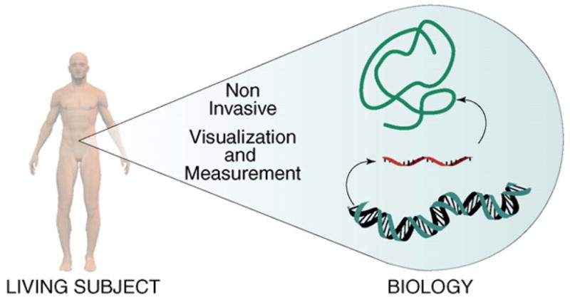
Molecular imaging permits non-invasive visualization and measurement of molecular and cell biology in living subjects, thereby complementing conventional anatomical imaging. Herein, we review the emerging application of molecular imaging for the study of musculoskeletal biology. Utilizing mainly bioluminescence and fluorescence techniques, molecular imaging has enabled in-vivo studies of (i) the activity of osteoblasts, osteoclasts, and hormones, (ii) the mechanisms of pathological cartilage and bone destruction, (iii) skeletal gene and cell therapy with and without biomaterial support, and (iv) the cellular processes in osteolysis and osteomyelitis. In these applications, musculoskeletal molecular imaging demonstrated feasibility for research in a myriad of musculoskeletal conditions ranging from bone fracture and arthritis to skeletal cancer. Importantly, these advances herald great potential for innovative clinical imaging in orthopedics, rheumatology, and oncology.
2009. Published by Elsevier Ltd.
Figures
Similar articles
-
[Analysis of Musculoskeletal Systems and Their Diseases. Musculoskeletal disease research by using imaging technique].Clin Calcium. 2015 Aug;25(8):1175-81. Clin Calcium. 2015. PMID: 26224675 Japanese.
-
[Homeostasis and Disorder of Musculoskeletal System.Cellular dynamics in musculoskeletal system visualized by intravital imaging techniques.].Clin Calcium. 2018;28(3):367-371. Clin Calcium. 2018. PMID: 29512528 Japanese.
-
Molecular imaging: an innovative force in musculoskeletal radiology.AJR Am J Roentgenol. 2013 Aug;201(2):264-77. doi: 10.2214/AJR.13.10713. Epub 2013 Jun 24. AJR Am J Roentgenol. 2013. PMID: 23795682 Review.
-
Qualitative and Quantitative Ultrashort Echo Time Imaging of Musculoskeletal Tissues.Semin Musculoskelet Radiol. 2015 Sep;19(4):375-86. doi: 10.1055/s-0035-1563733. Epub 2015 Nov 19. Semin Musculoskelet Radiol. 2015. PMID: 26583365 Review.
-
Fluorescence and Bioluminescence Imaging of Orthotopic Brain Tumors in Mice.Methods Mol Biol. 2017;1530:283-305. doi: 10.1007/978-1-4939-6646-2_17. Methods Mol Biol. 2017. PMID: 28150209
Cited by
-
Bioluminescent Imaging Systems for Assay Developments.Anal Sci. 2021 Feb 10;37(2):233-247. doi: 10.2116/analsci.20R003. Epub 2020 Sep 18. Anal Sci. 2021. PMID: 32963202 Review.
-
Imaging of alkaline phosphatase activity in bone tissue.PLoS One. 2011;6(7):e22608. doi: 10.1371/journal.pone.0022608. Epub 2011 Jul 25. PLoS One. 2011. PMID: 21799916 Free PMC article.
-
Molecular imaging of expression of vascular endothelial growth factor a (VEGF a) in femoral bone grafts transplanted into living mice.Cell Transplant. 2014;23(7):901-12. doi: 10.3727/096368912X667015. Epub 2013 Apr 12. Cell Transplant. 2014. PMID: 23582187 Free PMC article.
-
Combined in vivo optical and µCT imaging to monitor infection, inflammation, and bone anatomy in an orthopaedic implant infection in mice.J Vis Exp. 2014 Oct 16;(92):e51612. doi: 10.3791/51612. J Vis Exp. 2014. PMID: 25350287 Free PMC article.
-
Comparison of 3 real-time, quantitative murine models of staphylococcal biofilm infection by using in vivo bioluminescent imaging.Comp Med. 2014 Feb;64(1):25-33. Comp Med. 2014. PMID: 24512958 Free PMC article.
References
-
- Gross S, Piwnica-Worms D. Spying on cancer: molecular imaging in vivo with genetically encoded reporters. Cancer Cell. 2005;7:5–15. - PubMed
-
- Massoud TF, Gambhir SS. Molecular imaging in living subjects: seeing fundamental biological processes in a new light. Genes Dev. 2003;17:545–580. - PubMed
-
- Weissleder R, Mahmood U. Molecular imaging. Radiology. 2001;219:316–333. - PubMed
-
- Honigman A, et al. Imaging transgene expression in live animals. Mol Ther. 2001;4:239–249. - PubMed
Publication types
MeSH terms
Grants and funding
LinkOut - more resources
Full Text Sources
