Review
. 2010 Feb;20(2):79-91.
doi: 10.1016/j.tcb.2009.11.004. Epub 2010 Jan 4.

Intermediate filaments take the heat as stress proteins

Affiliations
Review

Intermediate filaments take the heat as stress proteins

D M Toivola et al. Trends Cell Biol. 2010 Feb.

Abstract

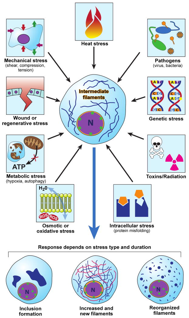
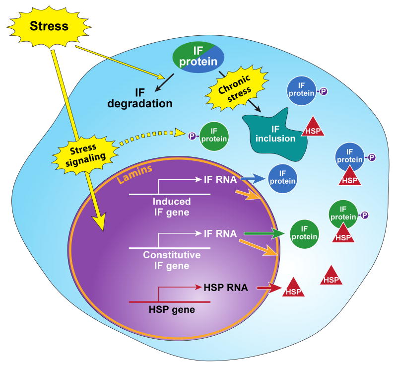
Intermediate filament (IF) proteins and heat shock proteins (HSPs) are large multimember families that share several features, including protein abundance, significant upregulation in response to a variety of stresses, cytoprotective functions, and the phenocopying of several human diseases after IF protein or HSP mutation. We are now coming to understand that these common elements point to IFs as important cellular stress proteins with some roles akin to those already well-characterized for HSPs. Unique functional roles for IFs include protection from mechanical stress, whereas HSPs are characteristically involved in protein folding and as chaperones. Shared IF and HSP cytoprotective roles include inhibition of apoptosis, organelle homeostasis, and scaffolding. In this report, we review data that corroborate the view that IFs function as highly specialized cytoskeletal stress proteins that promote cellular organization and homeostasis.


Figures

Fig.1
Fig.1. Multiple forms of stress modulate IFs
Cells are exposed to various forms of stress that originate from within or from the environment. Depending on the type and duration of stress encountered, IFs may respond by (a) forming cell-specific inclusions (e.g., Mallory-Denk bodies in alcoholic and nonalcoholic steatohepatitis [21, 84]); (b) up-regulating or de-novo generation of filaments (e.g. K19/K20 following pancreatic injury, [17]; or (c) reorganizing filaments (e.g. K18 following shear stress [56]).
Fig.2
Fig.2. Keratin IFs and HSPs are upregulated after organ-specific stress in the liver and pancreas
Livers from untreated mice or mice fed 0.1% 3,5-diethoxycarbonyl-1,4-dihydrocollidine (DDC) for 6 weeks (A, C) and, pancreata from untreated mice or mice injected hourly with 50 μg/kg caerulein 7 times, then recovered for 17 hours (B, D), were analyzed. (A-B) Total protein lysates from 3 different mice/treatment were probed with antibodies to K8, K18, K19, Hsp27, Hsp72, and tubulin (as a loading control). Alternatively, sections (1-2 μm) from fresh frozen livers (C) and pancreata (D) were fixed with cold acetone (10 minutes) then triple-stained to visualize K8/K18 (red), K19 (green), and nuclei (blue). Co-localization of K8/K18 and K19 appears yellow. Note the induction of K8, K18 and K19 (but not tubulin) upon liver stress (DDC; A, C) and pancreatic stress (caerulein; B, D). In the pancreas, K19 is dramatically upregulated upon recovery from caerulein-treatment (B) in acinar cells where it forms extensive cytoplasmic filaments with the resident K8/K18 filaments (Dd,f) compared to the preferential apico-lateral distribution (arrows) in untreated pancreata (Dc,e). In DDC-fed mice, hepatocyte K8/K18 upregulation (A) is reflected by the denser keratin networks (C) compared to control (Ca), while K19 upregulation is reflected by proliferation of K19-positive ductal cells (arrowheads, Cb). Scale bar in a for a-b is 50 m and in c for c-f is 20μm. This figure illustrates: (i) the upregulation of several keratin IFs in two different organs upon two unique and organ-specific toxic stresses; (ii) the joint upregulation of keratin IFs and two different HSPs during the two toxic treatments in the pancreas and liver, and (iii) that upregulation of the “stress protein” K19 is context dependent and may reflect either de-novo cytoplasmic expression (and overall increased expression) in acinar cells or hyperproliferation of ductal cells in the liver.
Fig.3
Fig.3. Specific stresses upregulate IFs and HSPs
Environmental or internal stresses (see Fig.1) initiate stress signaling cascades, which activate the stress response and transcriptional machineries that induce the expression of the classical stress-induced HSP genes (constitutive HSP genes are not shown). Stress also induces the transcription and translation of constitutive/resident IF genes (shown in green circles) nonresident IF genes (shown in blue circles) and the nuclear lamin IFs (shown in orange). The increased expression of IFs provides pools for new filament assembly (not shown), binding to IFAPs (exemplified by the established binding with HSPs, red triangles). IF posttranslational modifications (e.g., phosphorylation (P)), serve important functions in IF filament dynamics, binding to IFAPs and targeting for degradation initially or after recovery from stress. Exposure to chronic stress, in the appropriate context, leads to inclusion body formation which also include HSPs and other chaperones. These alterations in IF homeostasis and dynamics help promote the cellular response which allows cells to migrate, divide, differentiate, or die.

Similar articles

Cited by

References

    1. Morimoto RI, Santoro MG. Stress-inducible responses and heat shock proteins: new pharmacologic targets for cytoprotection. Nat Biotechnol. 1998;16(9):833–8. - PubMed
    1. Kampinga HH, et al. Guidelines for the nomenclature of the human heat shock proteins. Cell Stress Chaperones. 2009;14(1):105–11. - PMC - PubMed
    1. Akerfelt M, et al. Heat shock factors at a crossroad between stress and development. Ann N Y Acad Sci. 2007;1113:15–27. - PubMed
    1. Morimoto RI. Proteotoxic stress and inducible chaperone networks in neurodegenerative disease and aging. Genes Dev. 2008;22(11):1427–38. - PMC - PubMed
    1. van Noort JM. Stress proteins in CNS inflammation. J Pathol. 2008;214(2):267–75. - PubMed

Publication types

Substances