

Methamphetamine acts on subpopulations of neurons regulating sexual behavior in male rats
- PMID: 20045448
- PMCID: PMC2837118
- DOI: 10.1016/j.neuroscience.2009.12.070
Methamphetamine acts on subpopulations of neurons regulating sexual behavior in male rats
Abstract
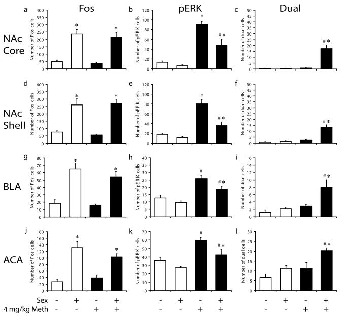
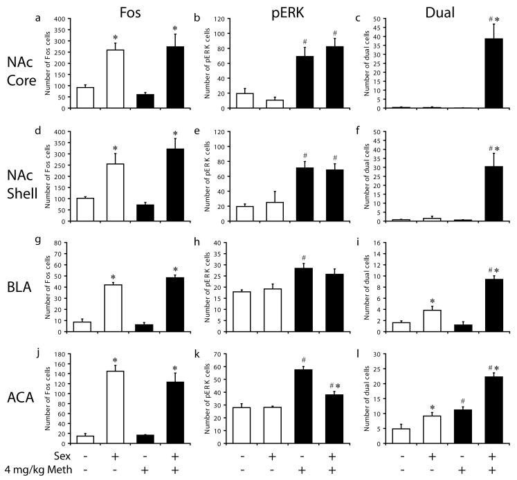
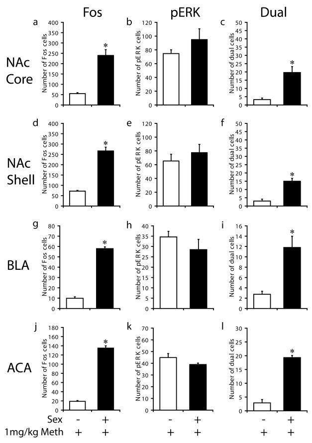
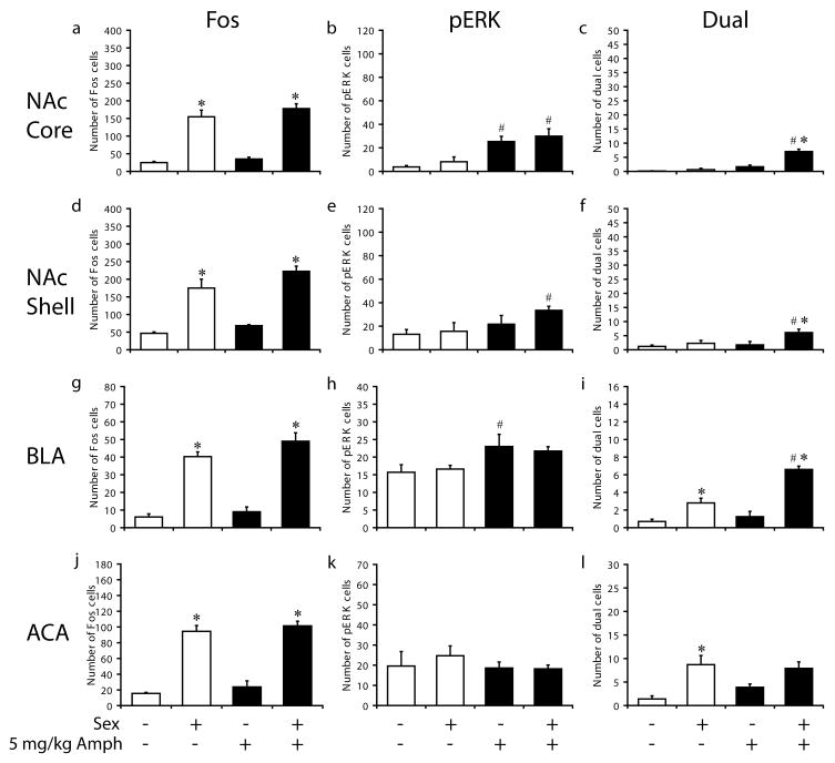
Methamphetamine (Meth) is a highly addictive stimulant. Meth abuse is commonly associated with the practice of sexual risk behavior and increased prevalence of Human Immunodeficiency Virus and Meth users report heightened sexual desire, arousal, and sexual pleasure. The biological basis for this drug-sex nexus is unknown. The current study demonstrates that Meth administration in male rats activates neurons in brain regions of the mesolimbic system that are involved in the regulation of sexual behavior. Specifically, Meth and mating co-activate cells in the nucleus accumbens core and shell, basolateral amygdala, and anterior cingulate cortex. These findings illustrate that in contrast to current belief drugs of abuse can activate the same cells as a natural reinforcer, that is sexual behavior, and in turn may influence compulsive seeking of this natural reward.
Keywords: addiction; basolateral amygdala; nucleus accumbens; prefrontal cortex; reproduction; substance abuse.
Copyright 2010 IBRO. Published by Elsevier Ltd. All rights reserved.
Figures

Similar articles
-
Maladaptive Sexual Behavior Following Concurrent Methamphetamine and Sexual Experience in Male Rats is Associated with Altered Neural Activity in Frontal Cortex.Neuropsychopharmacology. 2017 Sep;42(10):2011-2020. doi: 10.1038/npp.2017.1. Epub 2017 Jan 4. Neuropsychopharmacology. 2017. PMID: 28051103 Free PMC article.
-
Concurrent exposure to methamphetamine and sexual behavior enhances subsequent drug reward and causes compulsive sexual behavior in male rats.J Neurosci. 2011 Nov 9;31(45):16473-82. doi: 10.1523/JNEUROSCI.4013-11.2011. J Neurosci. 2011. PMID: 22072697 Free PMC article.
-
Oxytocin treatment in the prelimbic cortex reduces relapse to methamphetamine-seeking and is associated with reduced activity in the rostral nucleus accumbens core.Pharmacol Biochem Behav. 2019 Aug;183:64-71. doi: 10.1016/j.pbb.2019.06.002. Epub 2019 Jun 13. Pharmacol Biochem Behav. 2019. PMID: 31202809
-
"Sexy stimulants": the interaction between psychomotor stimulants and sexual behavior in the female brain.Pharmacol Biochem Behav. 2014 Jun;121:53-61. doi: 10.1016/j.pbb.2013.11.006. Epub 2013 Nov 20. Pharmacol Biochem Behav. 2014. PMID: 24269964 Review.
-
Sex differences in methamphetamine use disorder perused from pre-clinical and clinical studies: Potential therapeutic impacts.Neurosci Biobehav Rev. 2022 Jun;137:104674. doi: 10.1016/j.neubiorev.2022.104674. Epub 2022 Apr 20. Neurosci Biobehav Rev. 2022. PMID: 35452744 Free PMC article. Review.
Cited by
-
Drug-taking in a socio-sexual context enhances vulnerability for addiction in male rats.Neuropsychopharmacology. 2019 Feb;44(3):503-513. doi: 10.1038/s41386-018-0235-1. Epub 2018 Oct 6. Neuropsychopharmacology. 2019. PMID: 30337639 Free PMC article.
-
Cathinone Use Disorder in the Context of Slam Practice: New Pharmacological and Clinical Challenges.Front Psychiatry. 2020 Jul 22;11:705. doi: 10.3389/fpsyt.2020.00705. eCollection 2020. Front Psychiatry. 2020. PMID: 32792999 Free PMC article.
-
Within-animal comparisons of novelty and cocaine neuronal ensemble overlap in the nucleus accumbens and prefrontal cortex.Behav Brain Res. 2020 Feb 3;379:112275. doi: 10.1016/j.bbr.2019.112275. Epub 2019 Oct 12. Behav Brain Res. 2020. PMID: 31614186 Free PMC article.
-
The role of dopaminergic and serotonergic transmission in the processing of primary and monetary reward.Neuropsychopharmacology. 2020 Aug;45(9):1490-1497. doi: 10.1038/s41386-020-0702-3. Epub 2020 May 11. Neuropsychopharmacology. 2020. PMID: 32392573 Free PMC article.
-
Neurofunctional Differences Related to Methamphetamine and Sexual Cues in Men With Shorter and Longer Term Abstinence Methamphetamine Dependence.Int J Neuropsychopharmacol. 2020 Apr 21;23(3):135-145. doi: 10.1093/ijnp/pyz069. Int J Neuropsychopharmacol. 2020. PMID: 31995187 Free PMC article.
References
-
- Agmo A. Male rat sexual behavior. Brain Res Brain Res Protoc. 1997;1:203–209. - PubMed
-
- Agmo A, Berenfeld R. Reinforcing properties of ejaculation in the male rat: role of opioids and dopamine. Behav Neurosci. 1990;104:177–182. - PubMed
-
- Agmo A, Federman I, Navarro V, Padua M, Velazquez G. Reward and reinforcement produced by drinking water: Role of opioids and dopamine receptor subtypes. Pharmacol Biochem Behav. 1993;46 - PubMed
-
- Balfour ME, Yu L, Coolen LM. Sexual behavior and sex-associated environmental cues activate the mesolimbic system in male rats. Neuropsychopharmacology. 2004;29:718–730. - PubMed
-
- Baum MJ, Everitt BJ. Increased expression of c-fos in the medial preoptic area after mating in male rats: Role of afferent inputs from the medial amygdala and midbrain central tegmental field. Neuroscience. 1992;50:627–646. - PubMed
Publication types
MeSH terms
Substances
Grants and funding
LinkOut - more resources
Full Text Sources
Medical
