. 2010 Mar 15;339(2):519-27.
doi: 10.1016/j.ydbio.2009.12.030. Epub 2010 Jan 4.

Distinct enhancers at the Pax3 locus can function redundantly to regulate neural tube and neural crest expressions

Affiliations

Distinct enhancers at the Pax3 locus can function redundantly to regulate neural tube and neural crest expressions

Karl R Degenhardt et al. Dev Biol. .

Abstract

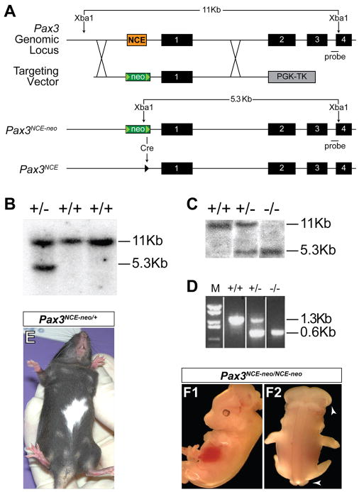
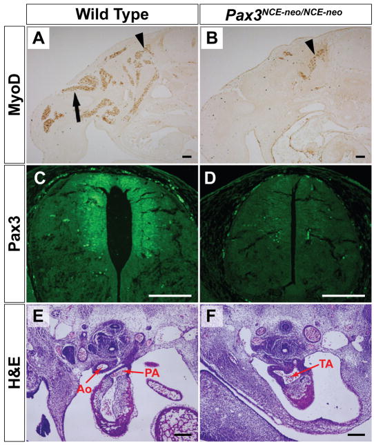
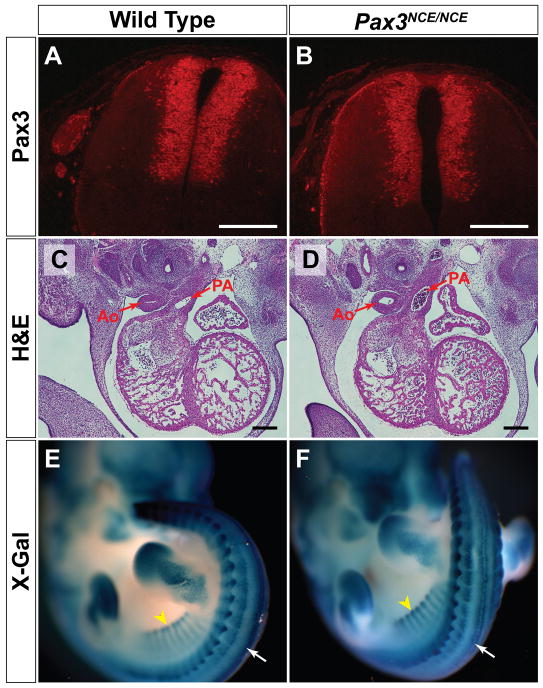
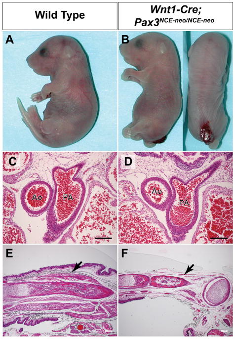
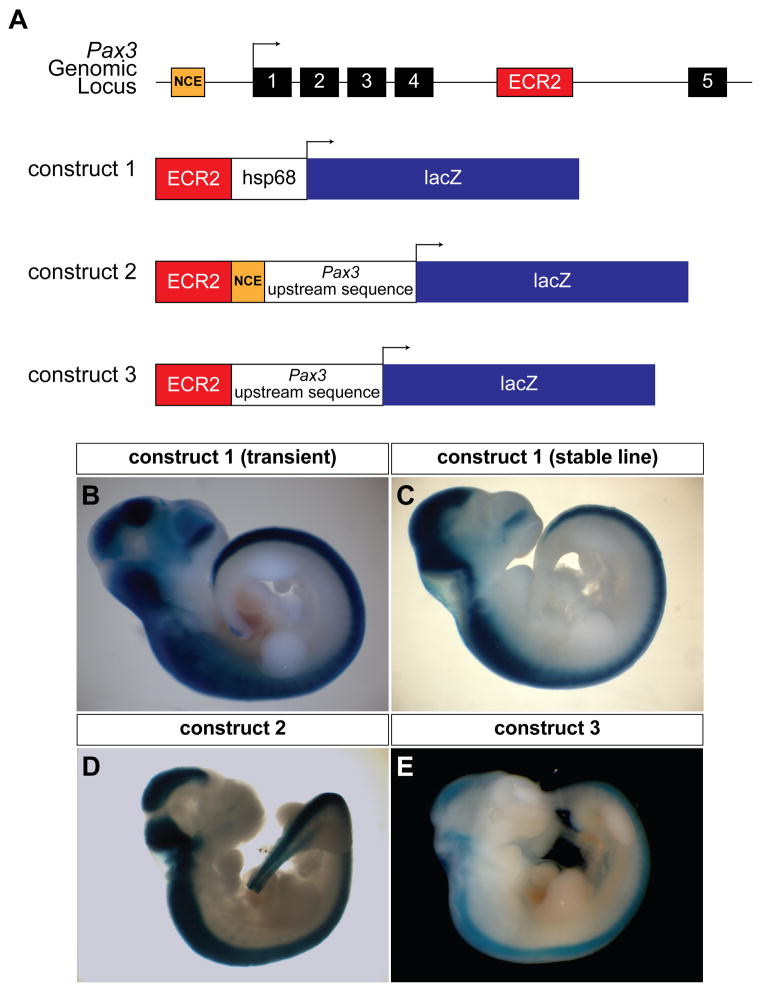
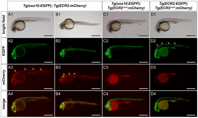
Pax3 is a transcription factor expressed in somitic mesoderm, dorsal neural tube and pre-migratory neural crest during embryonic development. We have previously identified cis-acting enhancer elements within the proximal upstream genomic region of Pax3 that are sufficient to direct functional expression of Pax3 in neural crest. These elements direct expression of a reporter gene to pre-migratory neural crest in transgenic mice, and transgenic expression of a Pax3 cDNA using these elements is sufficient to rescue neural crest development in mice otherwise lacking endogenous Pax3. We show here that deletion of these enhancer sequences by homologous recombination is insufficient to abrogate neural crest expression of Pax3 and results in viable mice. We identify a distinct enhancer in the fourth intron that is also capable of mediating neural crest expression in transgenic mice and zebrafish. Our analysis suggests the existence of functionally redundant neural crest enhancer modules for Pax3.


Figures

Figure 1
Figure 1. Targeted Deletion of the Pax3 upstream NCE
(A). Schematic representation of targeting strategy. The NCE (orange box) is replaced by a floxed neomycin resistance cassette (neo) to produce the Pax3NCE-neo allele after homologous recombination. Cre recombinase removes the neomycin cassette, leaving a single loxP site (green triangle), to produce Pax3NCE. (B, C). Confirmation of targeted ES cells (B) and mice (C) by Southern blot using a probe 3′ of the targeting arms. (D). After cre recombination, the removal of the NCE is confirmed by PCR. E. Pax3NCE-neo/+ mice have white belly spots. (F). Pax3NCE-neo/NCE-neo E13.5 embryos have neural tube defects (white arrows).
Figure 2
Figure 2. Pax3NCE-neo/NCE-neo embryos display cardiac and limb muscle defects at E12.5
(A, B). Immunohistochemistry with a MyoD antibody reveals hypaxial myoblasts (black arrow) that have appropriately migrated to the limb in the wild type embryo (A), while no MyoD positive cells are detected in the limb of the Pax3NCE-neo/NCE-neo mutant (B). Epaxial myoblasts expressing MyoD (arrowhead) are present. (C, D). Pax3 protein is expressed in the dorsal neural tube of wild type (C) but not Pax3NCE-neo/NCE-neo (D) embryos. (E, F). H&E staining reveals septation of the aorta (Ao) and pulmonary artery (PA) in wild type embryo (E), while the truncus arteriosus (TA) is unseptated in the Pax3NCE-neo/NCE-neo littermate (F). Scale bar = 200 microns.
Figure 3
Figure 3. Removal of the PGK-neo cassette restores Pax3 expression, and results in normal conotruncal anatomy and Pax3 transcriptional activity despite the loss of the NCE
(A, B). Immunohistochemistry reveals normal Pax3 expression in the dorsal neural tube of an E12.5 Pax3NCE/NCE embryo (B) as compared to wild type (A). (C, D). Both wild type (C) and Pax3NCE/NCE (D) mice have normal septation of the aorta (Ao) and pulmonary artery (PA) at E12.5. E, F. X-gal staining of P34TKZ Pax3 reporter mice at E11.5 reveals normal expression of β-galactosidase in the neural crest (white arrow) and somites (black arrowhead) in wild type (E) and Pax3NCE/NCE (F) mice. Scale bar = 200 microns.
Figure 4
Figure 4. Restoration of Pax3 expression in neural crest rescues embryonic development
(A, B). Wnt1-cre, Pax3NCE-neo/NCE-neo mice survive to birth, but occasionally display spinal bifida (B) not seen in control littermates (A). (C–F). H&E staining of newborns shows normal septation of the aorta (Ao) and pulmonary artery (PA) in both wild type (C) and Wnt1-cre, Pax3NCE-neo/NCE-neo mice (D), but severe deficiency of forelimb musculature in Wnt1-cre, Pax3NCE-neo/NCE-neo mice (arrow, F) as compared to wild type (arrow, E). Scale bar = 200 microns.
Figure 5
Figure 5. ECR2 directs neural crest expression in transgenic mouse embryos
(A). Schematic representation of the region of the Pax3 locus containing the NCE (orange box) and ECR2 (red box). Black boxes represent exons. Three transgene constructs are depicted using ECR2 with the hsp68 minimal promoter as well as Pax3 upstream sequence with and without the NCE. (B). E10.5 transient transgenic embryo showing strong dorsal neural tube expression throughout the anterior-posterior axis. (C). The same pattern is seen in the stable line with Construct 1. (D). Transient transgenic embryo with ECR2 and the 1.6 kb Pax3 upstream region shows robust dorsal neural tube expression. (E). Transient transgenic embryo showing dorsal neural tube expression despite the removal of the NCE from the Pax3 upstream sequence.
Figure 6
Figure 6. Analysis of transgenic zebrafish shows that ECR2 directs expression to the dorsal neural tube and that this expression requires Lef1/TCF sequences
(A1–A4). Lateral view of a 1 dpf zebrafish embryo that is stably transgenic for Sox10:EGPF and transiently transgenic for ECR2:mCherry demonstrates strong dorsal neural tube expression (yellow arrowheads). (B1–B4). Dorsal view of a similarly injected embryo as shown in column A. (C1–C4). Lateral view of a 24hpf zebrafish embryo that is stably transgenic for Sox10:EGPF and injected with an ECR2ΔLef:mCherry transgene shows that mutations in the Lef1/TCF sequences abrogates expression. (D1–D4). Lateral view of a 24 hpf zebrafish embryo that has been injected with both ECR2:EGFP and ECR2ΔLef:mCherry transgenes shows EGFP expression in the dorsal neural tube (yellow arrowheads). Despite effective transposase activity, no mCherry expression is detected. Scale bar = 500 microns.

References

    1. Antonellis A, Huynh JL, Lee-Lin SQ, Vinton RM, Renaud G, Loftus SK, Elliot G, Wolfsberg TG, Green ED, McCallion AS, Pavan WJ. Identification of neural crest and glial enhancers at the mouse Sox10 locus through transgenesis in zebrafish. PLoS Genet. 2008;4:e1000174. - PMC - PubMed
    1. Auerbach R. Analysis of the developmental effects of a lethal mutation in the house mouse. J Exper Zool. 1954;127:305–329.
    1. Bang AG, Papalopulu N, Goulding MD, Kintner C. Expression of Pax-3 in the lateral neural plate is dependent on a Wnt-mediated signal from posterior nonaxial mesoderm. Dev Biol. 1999;212:366–80. - PubMed
    1. Bang AG, Papalopulu N, Kintner C, Goulding MD. Expression of Pax-3 is initiated in the early neural plate by posteriorizing signals produced by the organizer and by posterior non-axial mesoderm. Development. 1997;124:2075–85. - PubMed
    1. Blackwood EM, Kadonaga JT. Going the distance: a current view of enhancer action. Science. 1998;281:60–3. - PubMed

Publication types

MeSH terms

Substances

LinkOut - more resources