. 2010 Mar 15;339(2):307-19.
doi: 10.1016/j.ydbio.2009.12.035. Epub 2010 Jan 4.

Mago Nashi, Tsunagi/Y14, and Ranshi form a complex that influences oocyte differentiation in Drosophila melanogaster

Affiliations

Mago Nashi, Tsunagi/Y14, and Ranshi form a complex that influences oocyte differentiation in Drosophila melanogaster

Jordan P Lewandowski et al. Dev Biol. .

Abstract

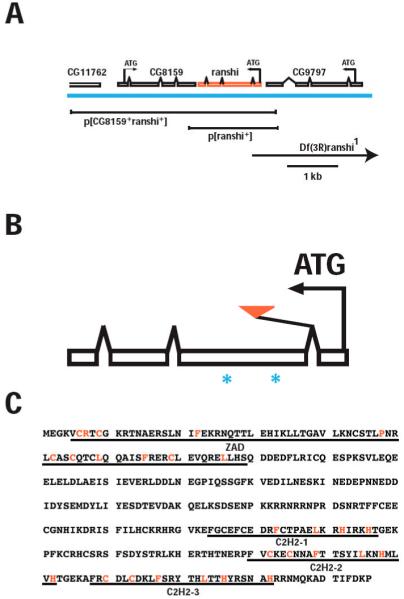
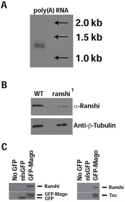
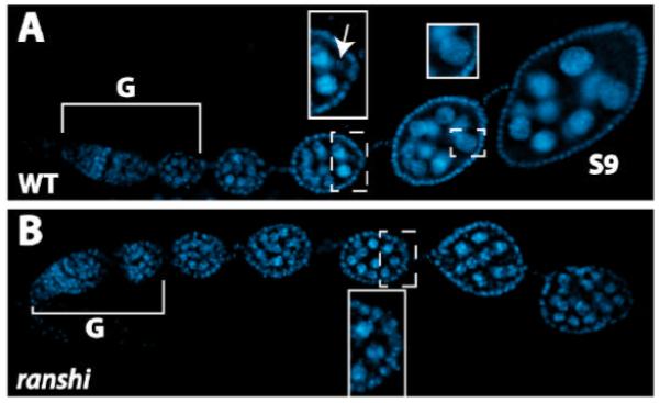
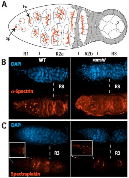
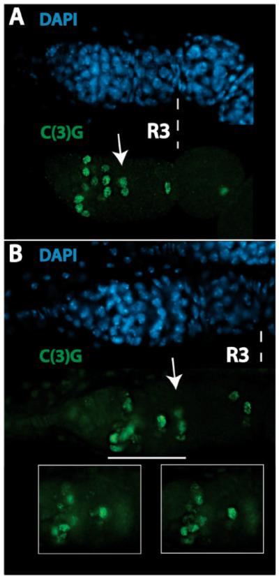
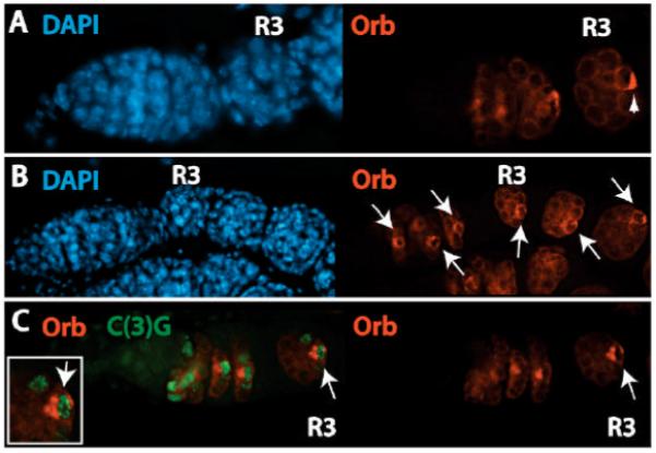
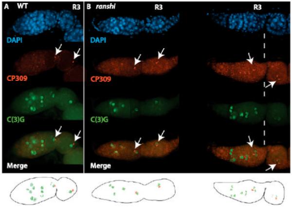
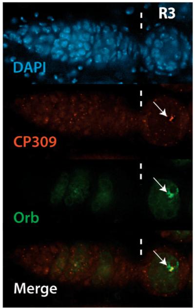
During Drosophila melanogaster oogenesis, a germline stem cell divides forming a cyst of 16 interconnected cells. One cell enters the oogenic pathway, and the remaining 15 differentiate as nurse cells. Although directed transport and localization of oocyte differentiation factors within the single cell are indispensible for selection, maintenance, and differentiation of the oocyte, the mechanisms regulating these events are poorly understood. Mago Nashi and Tsunagi/Y14, core components of the exon junction complex (a multiprotein complex assembled on spliced RNAs), are essential for restricting oocyte fate to a single cell and for localization of oskar mRNA. Here we provide evidence that Mago Nashi and Tsunagi/Y14 form an oogenic complex with Ranshi, a protein with a zinc finger-associated domain and zinc finger domains. Genetic analyses of ranshi reveal that (1) 16-cell cysts are formed, (2) two cells retain synaptonemal complexes, (3) all cells have endoreplicated DNA (as observed in nurse cells), and (4) oocyte-specific cytoplasmic markers accumulate and persist within a single cell but are not localized within the posterior pole of the presumptive oocyte. Our results indicate that Ranshi interacts with the exon junction complex to localize components essential for oocyte differentiation within the posterior pole of the presumptive oocyte.


Figures

Figure 1
Figure 1
Genomic organization of ranshi and amino acid sequence of Ranshi. (A) Genomic organization of ranshi showing its position relative to the flanking genes CG11762, CG8159 and CG9797. The centromere is to the left and telomere to the right. The blue horizontal line indicates genomic DNA and above the genomic DNA are predicted transcripts for the genes. For each gene the predicted translational start sites (ATG) and the direction of transcription (arrows) are indicated. Below the genomic DNA the rescuing transgenes and deletion (Df, indicated by the arrow to show that the deletion extends beyond the genomic region depicted) of the genomic region are illustrated. (B) The relative position of ranshi1 (red triangle) and of the regions chosen for generating peptides (*) employed for antibody production are shown. (C) The predicted Ranshi amino acid residues. The domains identified using Pfam to search databases are as follows: zing finger-associated motif (ZAD; amino acid residues 5-79) and the zinc-finger motifs in Ranshi are indicated (C2H2-1, C2H2-2 and C2H2-3).
Figure 2
Figure 2
The ranshi locus encodes a 1.3 kb RNA and an ~60 kDa protein that co-immunoprecipitates with Mago and Tsu/Y14. (A) RNA blot showing that a single ~1.3 kb transcript is detected in ovarian extracts of poly(A) RNA. (B) In ovarian extracts of protein isolated from wild type (WT), α-Ranshi identifies a single protein with an apparent Mr of ~60 kDa. However, in ovarian extracts of homozygous ranshi1 females the amount of Ranshi is reduced, indicating that the antibody is immunoreactive with Ranshi. (C) Immunoprecipitation with anti-GFP attached to beads preferentially immunoprecipitates GFP-Mago, Tsu/Y14 and Ranshi but not nlsGFP.
Figure 3
Figure 3
Ovaries of homozygous ranshi1 females contain 16-cell cysts blocked prior to stage 3 and 16 germline nuclei containing endoreplicated DNA. In both panels anterior is to the left. Brackets define germaria (G) and S9 indicates a stage 9 egg chamber. (A) DAPI staining of a wild-type ovariole. The posterior end of an egg chamber (anterior stippled rectangular box) is magnified in the rectangular box to illustrate the karyosome (indicated by the arrow). A nurse cell nucleus (posterior, stippled, square box) is magnified in the posterior square box to illustrate dispersed chromosomes. (B) DAPI stained ovarioles dissected from homozygous ranshi1 females contain 16-cell cysts and egg chambers without identifiable karyosomes. A posterior pole of an egg chamber is a magnified within the box to illustrate the absence of a karyosome and the presence of nuclei with polytene chromosomes.
Figure 4
Figure 4
Spectrosome/fusome development in ovaries of homozygous ranshi1 females is indistinguishable from wild type. In all panels, anterior is to the left. (A) Cartoon of a germarium. Spectrosomes (Sp) and fusomes (Fu) are illustrated in red. Vertical lines demarcate germarial regions 1 (R1), 2a (R2a), 2b (R2b) and 3 (R3). (B) Spectrosome/fusome development in wild-type (left) and homozygous ranshi1 females are similar. In both (B) and (C), R3 is to the right of the dotted vertical line. (C) The distribution of Spectraplakin in wild-type and homozygous ranshi1 germaria is indistinguishable. The boxes contain magnified photographs of the anterior tip of the germaria to show the presence of spectrosomes.
Figure 5
Figure 5
Restriction of meiosis to a single cell requires ranshi+ function. In all panels, anterior is to the left and R3 indicates germarial region 3. (A) Synaptonemal complex formation and meiotic progression in a wild-type germarium. The arrow identifies a R2b cyst with two adjacent cells (pro-oocytes) that are C(3)G positive. (B) In ranshi mutant germaria meiosis is not restricted to a single cell, as assessed by the presence of synaptonemal complexes in two adjacent cells within R3. The arrow identifies a R2b cyst with at least 3 adjacent cells with synaptonemal complexes. The white horizontal line delineates the germarial region magnified in the boxes and shows synaptonemal complexes within different focal planes.
Figure 6
Figure 6
Accumulation of Orb within a single cell of 16-cell cysts is independent of ranshi+ but posterior pole localization of Orb requires Ranshi. In all panels, anterior is to the left and (R3) is germarial region 3. (A) Orb distribution within a wild-type germarium. The arrowhead indicates posterior localization of Orb in R3. (B) In ranshi ovaries Orb is detected within a single cell within 16-cell cysts but is evenly distributed throughout the cytoplasm of the cell. Orb persists in post-germarial egg chambers (indicated by the arrows). (C) Although adjacent cells within ranshi germaria are C(3)G positive, only one cell contains Orb, indicated by the arrow and magnified in the inset.
Figure 7
Figure 7
Centrosomes accumulate within one cell of ranshi 16-cell cysts but fail to localize within the posterior pole of the cell. In both panels, anterior is to the left, α-CP309 is employed to detect centrosomes, α-C(3)G detects synaptonemal complexes, arrows point to centrosomes and R3. (A) The progression of synaptonemal complex formation and the distribution of centrosomes in a wild-type germarium. (B) In ranshi germaria, centrosomes accumulate within the anterior pole of a single cell but fail to translocate to the posterior pole. In ranshi cysts containing two adjacent cells with synaptonemal complexes, centrosomes accumulate in one of the two cells with synaptonemal complexes. Below are cartoons of the merged CP309 and C(3)G images for wild-type and ranshi germaria.
Figure 8
Figure 8
Orb and centrosomes accumulate within the same cell in ranshi egg chambers. Illustrated is a ranshi germarium with posterior to the left and vertical dashed lines marking germarial region 3 (R3). The arrows indicate centrosomes (red) labeled with α-309 and Orb (green) labeled with α-Orb accumulating in one cell of a ranshi stage 1 egg chamber, indicating that an oocyte is specified in ranshi ovaries.

Similar articles

Cited by

References

    1. al-Mukhtar KA, Webb AC. An ultrastructural study of primordial germ cells, oogonia and early oocytes in Xenopus laevis. J. Embryol. Exp. Morphol. 1971;26:195–217. - PubMed
    1. Bolivar J, Huynh JR, Lopez-Schier H, Gonzalez C, St Johnston D, Gonzalez-Reyes A. Centrosome migration into the Drosophila oocyte is independent of BicD and egl, and of the organisation of the microtubule cytoskeleton. Development. 2001;128:1889–1897. - PubMed
    1. Brayer KJ, Segal DJ. Keep your fingers off my DNA: protein-protein interactions mediated by C2H2 zinc finger domains. Cell Biochem. Biophys. 2008;50:111–131. - PubMed
    1. Brown RS. Zinc finger proteins: getting a grip on RNA. Curr. Opin. Struct. Biol. 2005;15:94–98. - PubMed
    1. Büning J. The Insect Ovary: Ultrastructure, Previtellogenic Growth and Evolution. Chapman & Hall; New York: 1994.

Publication types

LinkOut - more resources