. 2010 Mar 17;50(6):564-76.
doi: 10.1016/j.visres.2009.12.014. Epub 2010 Jan 4.

The effect of age on compensation for a negative lens and recovery from lens-induced myopia in tree shrews (Tupaia glis belangeri)

Affiliations

The effect of age on compensation for a negative lens and recovery from lens-induced myopia in tree shrews (Tupaia glis belangeri)

Thomas T Norton et al. Vision Res. .

Abstract

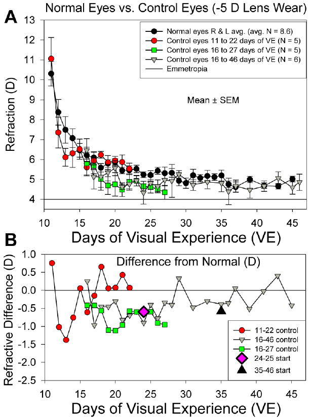
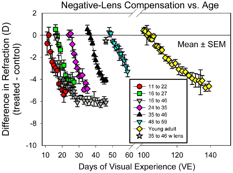
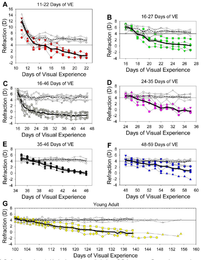
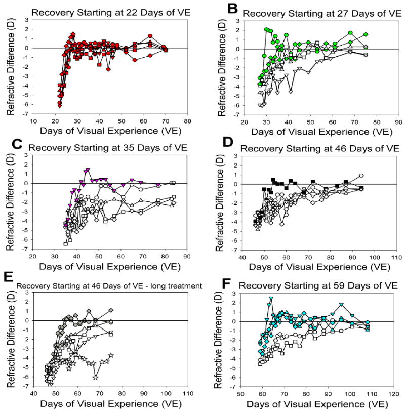
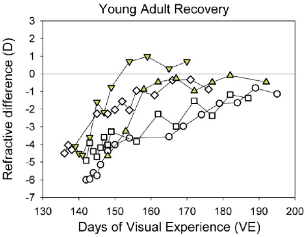
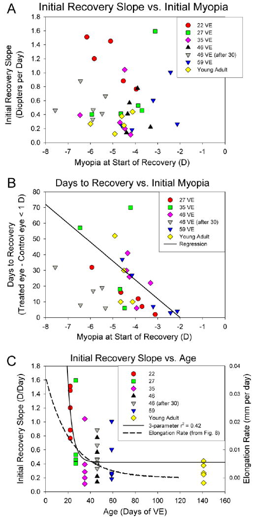
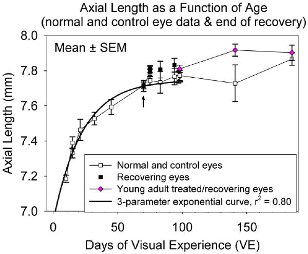
We examined in tree shrews the effect of age on the development of, and recovery from, myopia induced with a negative lens. Starting at 11, 16, 24, 35 or 48days after natural eye-opening (days of visual experience [VE]), juvenile tree shrews (n=5 per group) wore a monocular -5D lens for 11days. A long-term lens-wear group (n=6) began treatment at 16days of VE and wore the lens for 30days. A young adult group (n=5) began to wear a -5D lens between 93 and 107days of VE (mean+/-SD, 100+/-6days of VE) and wore the lens for 29-54days (mean+/-SD, 41.8+/-9.8days). The recovery phase in all groups was started by discontinuing -5D lens wear. Contralateral control eyes in the three youngest groups were compared with a group of age-matched normal eyes and showed a small (<1D), transient myopic shift. The amount of myopia that developed during lens wear was measured as the difference between the treated and control eye refractions. After 11days of lens wear, the induced myopia was similar for the four younger groups (near full compensation: 11days, -5.1+/-0.4D; 16days, -4.7+/-0.3D; 24days, -4.9+/-0.4D; 35days, -4.0+/-0.02) and slightly less in the oldest juvenile group (48days, -3.3+/-0.5D). The young adult animals developed -4.8+/-0.3D of myopia after a longer lens-wear period. The rate of compensation (D/day) was high in the 4 youngest groups and decreased in the 48-day and young adult groups. The refractions of the long-term lens-wear juvenile group remained stable after compensating for the -5D lens. During recovery, all animals in the youngest group recovered fully (<1D residual myopia) within 7days. Examples of both rapid (<10days) and slow recovery (>12days) occurred in all age groups except the youngest. Every animal showed more rapid recovery (higher recovery slope) in the first 4days than afterward. One animal showed extremely slow recovery. Based on the time-course of myopia development observed in the youngest age groups, the start of the susceptible period for negative-lens wear is around 11-15days after eye opening; the rate of compensation remains high until approximately 35days of VE and then gradually declines. Compensation is stable with continued lens wear. The emmetropization mechanism, both for lens compensation and recovery, remains active into young adulthood. The time-course of recovery is more variable than that of compensation and seems to vary with age, with the amount of myopia (weakly) and with the individual animal.


Figures

Fig. 1
Fig. 1
Experimental groups. Filled bars indicate the ages at which each group wore a monocular −5 D lens. “Rec” indicates the recovery period of unrestricted visual experience after lens wear was discontinued. Because the young adult group varied in the onset, length of lens wear, and length of the recovery period, the means and standard deviations are shown in the figure.
Fig. 2
Fig. 2
A. Normal- and control-eye refractive values (mean ± SEM) as a function of age. The control eyes from groups that began lens wear at 11 or 16 days of VE developed a small, transient myopic shift. Estimated emmetropia is an autorefractor value of +4 D due to the “small eye artifact” (Glickstein & Millodot, 1970; Ramamirtham et al., 2003; Norton et al., 2003). B. Average difference between the control-eye and normal-eye refractions for the three groups whose control eyes showed a decrease in refraction from normal. Starting differences (relative to normal eyes) for the 24- and 35-day control eyes are also shown.
Fig. 3
Fig. 3
Development of myopia during −5 D lens wear. Group mean ± SEM values of the refractive difference between the −5 D lens-wearing eyes and control eyes in the five juvenile groups with 11 days of treatment, the juvenile group with 30 days of lens treatment and the young adult group. The star at 46 days of VE indicates the with-the-lens refractive difference of the treated eyes of the long-term lens-wear group at the end of treatment.
Fig. 4
Fig. 4
Refractive values (with the lens removed) during −5 D lens wear. For each group, the treated eye and control eye refractions are shown, along with the mean refractions. Filled symbols, treated eyes; open symbols, control eyes; thick solid line, treated eye average; thick dotted line, control eye average. The abscissa for the young adult animals has been expanded to reflect the longer period of lens wear. Starting day for all animals in the Young Adult group were set to 100 days of VE.
Fig. 5
Fig. 5
Recovery of the treated eyes of individual juvenile animals starting at different ages is shown as the non-cycloplegic difference in refraction of the recovering eye and its fellow control eye. Filled symbols indicate animals that recovered quickly (10 days or less). The scale on the abscissa is the same in all graphs to facilitate comparisons of the rate of recovery.
Fig. 6
Fig. 6
Recovery of young adult animals, showing the refractive difference between the recovering and fellow control eyes for each of the five animals. Filled symbols indicate animals that recovered quickly (less than 10 days).
Fig. 7
Fig. 7
A. Initial recovery slope for all age groups as a function of the initial amount of myopia. This slope was unrelated to the amount of myopia that had developed. B. Excluding the youngest age group, the length of time to recover from the induced myopia (to < 1D) was weakly, but significantly related to the amount of myopia (r2 = 0.14, p < 0.05). The regression (solid line) includes the animal that was very slow to recover (projected recovery at 201 days), but that data point is not plotted on the graph. C. Initial recovery slope as a function of age. The solid line is a 3-parameter exponential function fitted to the initial recovery slopes. The dashed line is the daily rate of axial elongation calculated from the exponential function fitted to the normal axial lengths in Fig. 8.
Fig. 8
Fig. 8
Axial length of eyes (group mean ± SEM) as a function of age and treatment. Open squares indicate normal eyes (right and left eyes averaged) at the time of pedestal installation and control eyes at the end of recovery. Filled squares indicate recovering eyes at the end of the recovery period. The arrow indicates the post-recovery treated-eye axial lengths of the youngest group, which obscure the control eye values. Diamonds indicate the treated eyes of the young adult group before treatment, after -5 D lens wear and at the end of the recovery period. Adult data are plotted at the mean time of pedestal installation, end of treatment and end of recovery. The solid line is a 3-parameter exponential function fitted to the data from the normal (pre-treatment) measurements.

Similar articles

Cited by

References

    1. Diether S, Schaeffel F, Lambrou GN, Fritsch C, Trendelenburg AU. Effects of intravitreally and intraperitoneally injected atropine on two types of experimental myopia in chicken. Exp Eye Res. 2007;84:266–274. - PubMed
    1. Glickstein M, Millodot M. Retinoscopy and eye size. Science. 1970;168:605–606. - PubMed
    1. Graham B, Judge SJ. The effects of spectacle wear in infancy on eye growth and refractive error in the marmoset (Callithrix jacchus) Vision Res. 1999;39:189–206. - PubMed
    1. Green DG, Powers MK, Banks MS. Depth of focus, eye size and visual acuity. Vision Res. 1980;20:827–835. - PubMed
    1. Gwiazda J, Hyman L, Hussein M, Everett D, Norton TT, Kurtz D, Leske MC, Manny R, Marsh-Tootle W, Scheiman M. A randomized clinical trial of progressive addition lenses versus single vision lenses on the progression of myopia in children. Invest Ophthalmol Vis Sci. 2003;44:1492–1500. - PubMed

Publication types