. 2010 May;71(1):62-73.
doi: 10.1016/j.pep.2009.12.013. Epub 2010 Jan 4.

Recombinant derivatives of botulinum neurotoxin A engineered for trafficking studies and neuronal delivery

Affiliations

Recombinant derivatives of botulinum neurotoxin A engineered for trafficking studies and neuronal delivery

Philip A Band et al. Protein Expr Purif. 2010 May.

Abstract

Work from multiple laboratories has clarified how the structural domains of botulinum neurotoxin A (BoNT/A) disable neuronal exocytosis, but important questions remain unanswered. Because BoNT/A intoxication disables its own uptake, light chain (LC) does not accumulate in neurons at detectable levels. We have therefore designed, expressed and purified a series of BoNT/A atoxic derivatives (ad) that retain the wild type features required for native trafficking. BoNT/A1ad(ek) and BoNT/A1ad(tev) are full length derivatives rendered atoxic through double point mutations in the LC protease (E(224)>A; Y(366)>A). DeltaLC-peptide-BoNT/A(tev) and DeltaLC-GFP-BoNT/A(tev) are derivatives wherein the catalytic portion of the LC is replaced with a short peptide or with GFP plus the peptide. In all four derivatives, we have fused the S6 peptide sequence GDSLSWLLRLLN to the N-terminus of the proteins to enable site-specific attachment of cargo using Sfp phosphopantetheinyl transferase. Cargo can be attached in a manner that provides a homogeneous derivative population rather than a polydisperse mixture of singly and multiply-labeled molecular species. All four derivatives contain an introduced cleavage site for conversion into disulfide-bonded heterodimers. These constructs were expressed in a baculovirus system and the proteins were secreted into culture medium and purified to homogeneity in yields ranging from 1 to 30 mg per liter. These derivatives provide unique tools to study toxin trafficking in vivo, and to assess how the structure of cargo linked to the heavy chain (HC) influences delivery to the neuronal cytosol. Moreover, they create the potential to engineer BoNT-based molecular vehicles that can target therapeutic agents to the neuronal cytoplasm.


Figures

Figure 1
Figure 1. Amino acid sequence and features of the expressed BoNT/A derivatives in comparison with wt
Spaces between arrowheads and arrow tails represent regions of sequence identity omitted for simplicity. Numbers in the upper row correspond to amino acid residues in wt BoNT/A. Residues that are identical in all proteins are shown in black. Introduced mutations are shown in red. Added amino acids are shown in blue. Signal peptide required for insect cell secretion of the expressed derivatives into medium indicated by “SP” on blue background. Tags used for affinity chromatography are indicated: polyhistidine tag is indicated by “10 His” on a gray background; StrepTag II is indicated by “StrepTag II” on a purple background. “TEV” and amino acid sequence on a yellow background represent tobacco etch virus protease recognition sequence. Amino acids on an orange background represent enterokinase recognition sequence. “S6” on a pink background identifies a peptide tag used for site-specific attachment of cargo to the expressed proteins. “GFP” on a green background represents a portion of green fluorescent protein. The five proteins are aligned to illustrate homology between respective structural domains. Gaps have been introduced to facilitate the alignment. Spaces between rectangle-enclosed sequences represent sites of proteolytic cleavage. The disulfide bridges between residues of the light and heavy chains are indicated by horizontal brackets.
Figure 2
Figure 2. BoNT/Aadek propeptide purification
Reduced 12% SDS PAGE stained with Coomassie BB R-250. Panel A: Talon® chromatography purification: lane 1, wt BoNT/A, control; lane 2, unfractionated sample of concentrated and dialyzed Sf-900 II medium containing secreted propeptide BoNT/Aadek prior to loading on column; lane 3, column flow through; lane 4, wash 1, loading buffer; lane 5, wash 2, loading buffer with 20 mM imidazole; lane 6, elution, loading buffer with 200 mM imidazole. Panel B: StrepTactin agarose chromatography: lane 1, sample of concentrated and dialyzed fraction from Panel A, lane 6 prior to loading on column; lane 2, flow through; lanes 3 - 7, washes with loading buffer; lanes 8 - 12, elutions with loading buffer and 3mM desthiobiotin; lane 13, wt BoNT/A, control.
Figure 3
Figure 3. Processing of BoNT/Aadek propeptide
Panels A1, A2: Processing to heterodimer by proteolytic cleavage with recombinant enterokinase (rEK). One microgram of BoNT/Aadek propeptide per lane was treated with 0 - 5 U rEK at 16°C for 12 hours, separated by 12% SDS PAGE, and stained with Coomassie BB R-250. Panel A1: non-reduced samples. Panel A2: samples reduced by addition of β-mercaptoethanol. Lane 1, no rEK; lane 2, 0.001 U rEK; lane 3, 0.01 U rEK; lane 4, 0.1 U rEK; lane 5, 1 U rEK; lane 6, 5 U rEK; lane 7, wt BoNT/A, control. Panel B: Removal of 10-His tag from BoNT/Aadek propeptide by treatment with AcTEV. BoNT/Aadek was either treated with buffer (odd lane numbers), or treated with AcTEV protease (even lane numbers; 1 U per microgram, 30°C; see Methods for details) for the times indicated: lanes 1 and 2, one hour; lanes 3 and 4, two hours; lanes 5 and 6, three hours; lanes 7 and 8, four hours; lanes 9 and 10, six hours. Samples were loaded on a 12% SDS PAGE in the presence of β-mercaptoethanol, separated and transferred to nitrocellulose. Western blot was probed with HRP-coupled anti-His MAb (Santa Cruz, H-3 His probe, Cat # sc-8036 HRP). Note: The low MW band (approximately 30 kDa) in even lanes represents AcTEV protease which was supplied by Invitrogen as a 6-His tagged recombinant enzyme.
Figure 4
Figure 4. Structural analysis of tryptic peptides isolated from BoNT/Aadek
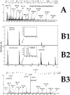
Panel A: ESI Q-TOF MS/MS spectrum of the C-terminal tryptic peptide isolated from enterokinase-processed BoNT/Aadek LC under reducing conditions. The b and y ion series have been included at the top of the figure to identify the peptide fragment peaks found in the spectrum, where the position of vertical bars separating each amino acid(s) correspond to the position of the respective ion m/z within the spectrum. For clarity, only the most intense peaks from y series have been labeled. Panels B1-B3: MALDI-TOF mass spectra and ESI Q-TOF MS/MS spectrum of the tryptic dipeptide with an internal disulfide bridge linking light and heavy chains of enterokinase-processed BoNT/Aadek. Panel B1: MALDITOF mass spectrum of an in-gel tryptic digest of the enterokinase-processed BoNT/Aadek LC, separated and isolated from a reduced SDS PAGE; Panel B2: MALDI-TOF mass spectrum of an in-gel tryptic digest of the enterokinase-processed BoNT/Aadek separated and isolated from an unreduced SDS PAGE. A peak at m/z 1489.84, matched thepredicted m/z of the dipeptide with internal disulfide bridge; Panel B3: ESI Q-TOF MS/MS spectrum of the dipeptide with m/z 1489.8 shown in Panel B2, confirming presence of the S-S bond in the dipeptide. The b and y ion series have been included at the top of the panel to identify the peptide fragment peaks found in the spectrum, where the position of vertical bars separating each amino acid(s) correspond to position of the respective ion m/z within the spectrum. Only the most intense peaks have been labeled for clarity.
Figure 5
Figure 5. In vitro fluorescent labeling of AcTEV-treated and rEK-treated BoNT/Aadek with Sfp phosphopantetheinyl transferase and CoA 547
Lanes 1 - 4, unreduced samples; lanes 5 - 8, samples reduced by addition of β-mercaptoethanol. Lanes 1, 3, 5, 7: 0.02 μg BoNT/Aadek; lanes 2, 4, 6, 8: 0.1 μg BoNT/Aadek. Panel A: 10.5 - 14% Criterion gel (Bio-Rad) stained with Bio-Safe Coomassie (Bio-Rad). Panel B: Western blot of gel shown in panel A scanned on a Typhoon 9500 scanner (GE Healthcare) using 300V PMT, 532/580 nm excitation/emission filter set (green).
Figure 6
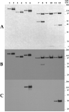
Figure 6. BoNT/Aadtev, ΔLC-Peptide-BoNT/Atev, and ΔLC-GFP-BoNT/Atev expressed in the baculovirus system, purified by metal chelate and StrepTactin affinity chromatography, and processed with AcTEV
Lanes 1 - 6, unreduced samples; lanes 7 - 12 samples reduced by addition of β-mercaptoethanol. Lanes 1, 2, 7, 8: BoNT/Aadtev; lanes 3, 4, 9, 10: ΔLC-Peptide-BoNT/Atev; lanes 5, 6, 11, 12: ΔLC-GFP-BoNT/Atev. Panel A: 10.5 - 14 % Criterion gel (Bio-Rad) stained with Bio-Safe Coomassie (Bio-Rad); odd lanes: 0.3 μg samples; even lanes: 1.0 μg samples. Panel B: Western blot probed with polyclonal antibody Pol001 raised against BoNT/A holotoxoid (Staten Serum Institut, Denmark); odd lanes: 3 ng samples; even lanes: 10 ng samples. Panel C: Western blot probed with monoclonal antibody against GFP (Clontech); odd lanes: 3 ng samples; even lanes: 10 ng samples.

Similar articles

Cited by

References

    1. National Institute of Occupational Safety and Health . Registry of Toxic Effects of Chemical Substances (R-TECS) National Institute of Occupational Safety and Health; Cincinnati, Ohio: 1996.
    1. Smith TJ, Lou J, Geren IN, Forsyth CM, Tsai R, Laporte SL, Tepp WH, Bradshaw M, Johnson EA, Smith LA, Marks JD. Sequence variation within botulinum neurotoxin serotypes impacts antibody binding and neutralization. Infect. Immun. 2005;73(9):5450–5457. - PMC - PubMed
    1. Simpson LL. Identification of the major steps in botulinum toxin action. Annu. Rev. Pharmacol. Toxicol. 2004;44:167–193. - PubMed
    1. Koriazova LK, Montal M. Translocation of botulinum neurotoxin light chain protease through the heavy chain channel. Nat. Struct. Biol. 2003;10(1):13–18. - PubMed
    1. Dekleva ML, DasGupta BR, Sathyamoorthy V. Botulinum neurotoxin type A radiolabeled at either the light or the heavy chain. Arch. Biochem. Biophys. 1989;274(1):235–240. - PubMed

Publication types

MeSH terms