. 2010 Jul;6(7):2740-50.
doi: 10.1016/j.actbio.2009.12.052. Epub 2010 Jan 4.

Functional biomimetic analogs help remineralize apatite-depleted demineralized resin-infiltrated dentin via a bottom-up approach

Affiliations

Functional biomimetic analogs help remineralize apatite-depleted demineralized resin-infiltrated dentin via a bottom-up approach

Jongryul Kim et al. Acta Biomater. 2010 Jul.

Abstract

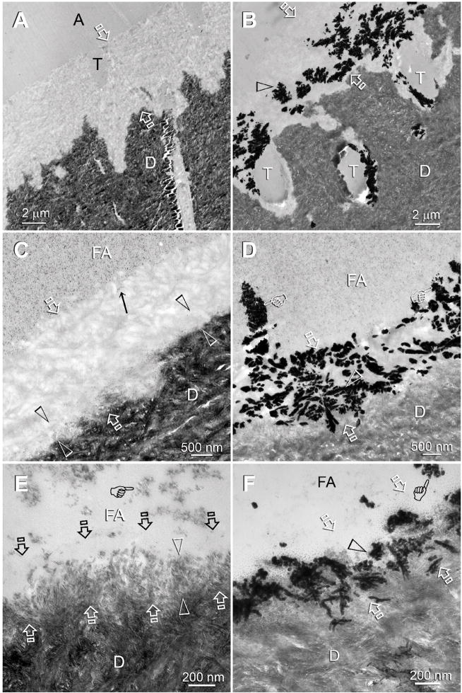
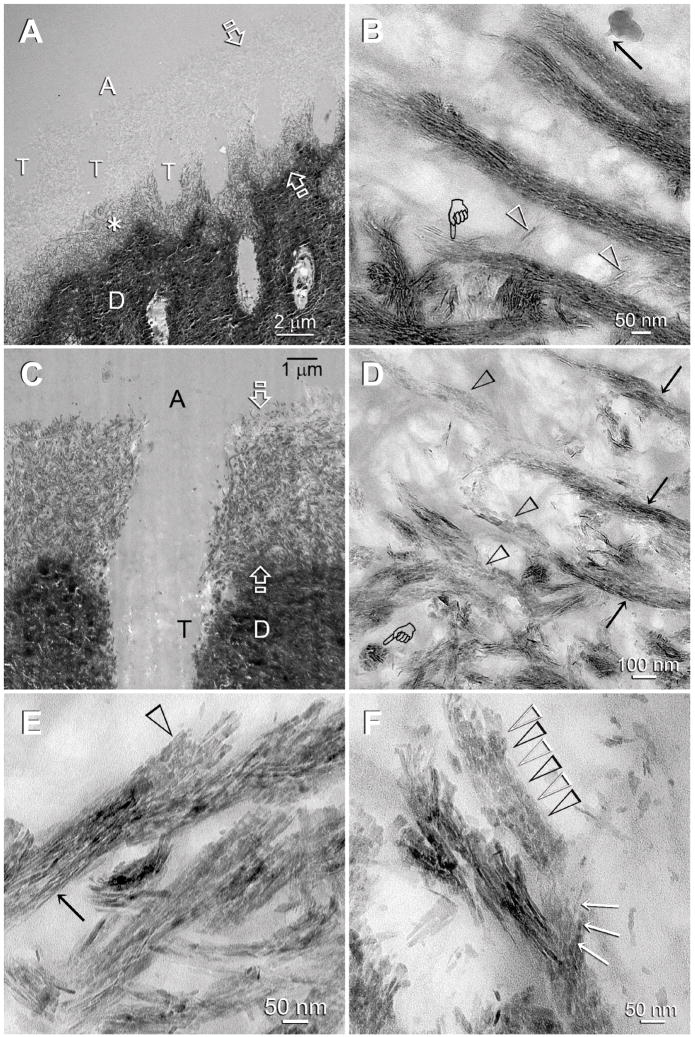
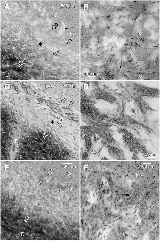
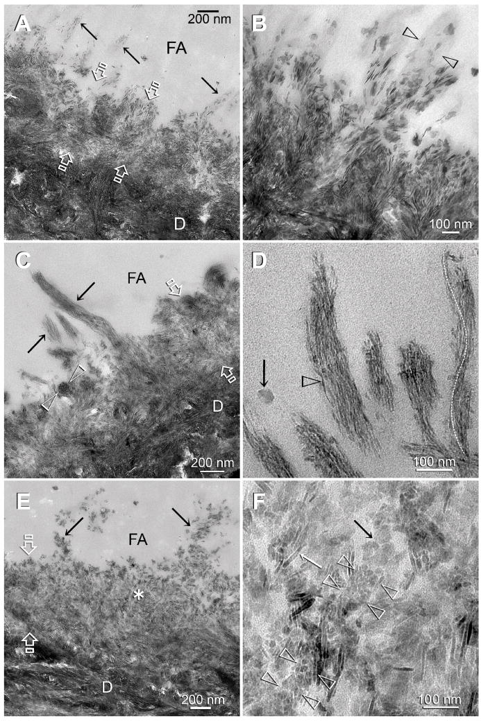
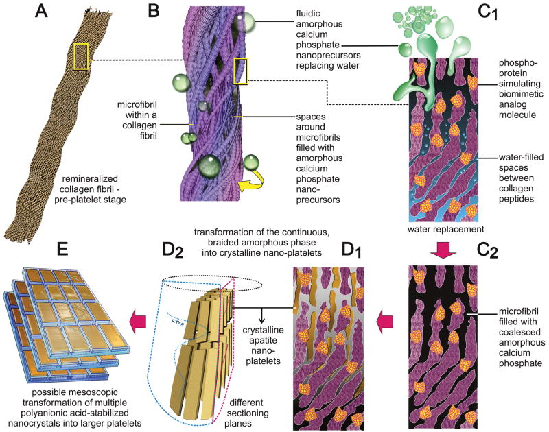
Natural biominerals are formed through metastable amorphous precursor phases via a bottom-up, nanoparticle-mediated mineralization mechanism. Using an acid-etched human dentin model to create a layer of completely demineralized collagen matrix, a bio-inspired mineralization scheme has been developed based on the use of dual biomimetic analogs. These analogs help to sequester fluidic amorphous calcium phosphate nanoprecursors and function as templates for guiding homogeneous apatite nucleation within the collagen fibrils. By adopting this scheme for remineralizing adhesive resin-bonded, completely demineralized dentin, we have been able to redeposit intrafibrillar and extrafibrillar apatites in completely demineralized collagen matrices that are imperfectly infiltrated by resins. This study utilizes a spectrum of completely and partially demineralized dentin collagen matrices to further validate the necessity for using a biomimetic analog-containing medium for remineralizing resin-infiltrated partially demineralized collagen matrices in which remnant seed crystallites are present. In control specimens in which biomimetic analogs are absent from the remineralization medium, remineralization could only be seen in partially demineralized collagen matrices, probably by epitaxial growth via a top-down crystallization approach. Conversely, in the presence of biomimetic analogs in the remineralization medium, intrafibrillar remineralization of completely demineralized collagen matrices via a bottom-up crystallization mechanism can additionally be identified. The latter is characterized by the transition of intrafibrillar minerals from an inchoate state of continuously braided microfibrillar electron-dense amorphous strands to discrete nanocrystals, and ultimately into larger crystalline platelets within the collagen fibrils. Biomimetic remineralization via dual biomimetic analogs has the potential to be translated into a functional delivery system for salvaging failing resin-dentin bonds.


Figures

Figure 1
Figure 1
TEM images of specimens that had not been subjected to biomimetic remineralization. These images provided baseline information on the depth of demineralization and extent of apatite dissolution in human dentin bonded with the three self-etch adhesives. Generic abbreviations - A: unfilled adhesive; FA: filled adhesive; D: dentin; T: dentinal tubule; Between open arrows: zone of demineralized dentin that was simultaneously infiltrated by the self-etch adhesive to produce an interdiffusion zone. A. Adper Prompt, the most aggressive adhesive, created a 5 Δm thick layer of completely-emineralized dentin. B. Adper Prompt-bonded dentin that had been immersed in a silver nitrate tracer solution. The electron dense silver deposits (open arrowhead) revealed water-rich, resin-sparse regions within the interdiffusion zone. C. Adper Scotchbond SE, a moderately aggressiveness adhesive, created a 2 Δm thick interdiffusion zone. A thin, partially-demineralized region (between open arrowheads) could be identified along the base of the interdiffusion zone. D. The corresponding silver impregnated section of Adper Scotchbond SE showing water-rich, resin-sparse regions (open arrowhead) within the interdiffusion zone. Some of the water channels (pointers) extended vertically from the dentin surface into the filled adhesive. E. Adper Easy Bond, the mildest of the three self-etch adhesives, created a 500 nm thick interdiffusion zone with a clearly discernible 300 nm thick partially-demineralized region along its base (between open arrowheads). Although the surface 200 nm part of the interdiffusion zone appeared completely-demineralized at this magnification, very fine remnant apatites could be identified at higher magnification. F. The corresponding silver impregnated section of Adper Easy Bond showing water channels (open arrowhead) within the interdiffusion zone and in the filled adhesive (pointer).
Figure 2
Figure 2
TEM images of experimental Adper Prompt specimens that had undergone biomimetic remineralization. The completely-demineralized resin infiltrated collagen matrices of the control specimens did not remineralize in the absence of biomimetic analogs (not shown). A: adhesive; D: dentin; T: dentinal tubule. A. A representative example of a specimen that exhibited features of an early stage of biomimetic remineralization. Partial remineralization (asterisk) occurred along the base of the original zone of completely-demineralized dentin (between open arrows). B. A high magnification view of Fig. 2A. Intrafibrillar remineralization could be seen in the form of an electron-dense, continuous ribbon-like mineral phase (pointer) that extended along the longitudinal axis of the collagen fibrils, accentuating the braided microfibrillar architecture of those remineralized fibrils. An electron-dense, amorphous droplet-like structure could be seen with an appendage extending into a remineralized collagen fibril (arrow). Individual intrafibrillar mineral platelets could not be identified at this stage. Extrafibrillar remineralization was minimal as the spaces between the collagen fibrils were filled with adhesive resins, and appeared in the form of needle-shaped crystallites (open arrowheads). C. A representative example of a specimen that exhibited features of a more mature stage of biomimetic remineralization. Intrafibrillar remineralization could be seen almost within the entire width of the original demineralized collagen matrix (between open arrows). D. A moderately high magnification view of Fig. 2C showing transformation of the continuous braided mineral phase (arrows) into discrete platelets (open arrowheads) within the same collagen fibrils. A transverse section through a remineralized fibril (pointer) clearly showed that it was fully filled with intrafibrillar minerals. E. A high magnification view, taken from a different specimen, illustrating the transition of the continuous braided mineral phase (arrow) into discrete platelets (open arrowhead) within a remineralized collagen fibril. This is the first time such a transformation has ever been demonstrated in situ within the same fibril (see also Fig. 3D). F. In this high magnification view, the remineralized fibril at the bottom still retained some of the continuous braided mineral phase (arrows), while the mineral phase within the fibril at the top had been transformed completely into discrete platelets (open arrowheads).
Figure 3
Figure 3
TEM images of control (A–B) and experimental (C–F) dentin specimens bonded with Adper Scotchbond SE. FA: filled adhesive; D: dentin; T: dentinal tubule; Between open arrows: the original 2Δm thick, partially demineralized interdiffusion zone. A. A control specimen that was retrieved after immersion in the Ca2+ and PO43−-containing simulated body fluid (SBF) for the entire experimental period. There was an increase in the thickness and density of the partially-demineralized region within the interdiffusion zone, probably due to epitaxial growth over remnant seed crystallites (see Fig. 1C). However, in the absence of biomimetic analogs, there was no remineralization of the surface, completely-demineralized part of the interdiffusion zone (between open arrowheads). The surface collagen fibrils (arrow) were electron lucent (i.e. devoid of minerals). B. A high magnification view of the location marked by the asterisk in Fig. 3A. Remineralization was predominantly extrafibrillar along the surface of the collagen fibrils (between open arrowheads). Intrafibrillar remineralization could not be recognized, probably due to depletion of remnant intrafibrillar seed crystallites from this location. C. An experimental specimen that exhibited features of an early stage of biomimetic remineralization. Unlike the control specimen (Fig. 3A), the surface part of the interdiffusion zone (between open arrowheads) was remineralized when biomimetic analogs are included in the SBF. The mode of remineralization was intrafibrillar in nature and consisted of the electron dense braided mineral phase that at this stage appeared completely different from the mineral phase present in the underlying interdiffusion zone (asterisk). D. A high magnification view of the electron dense braided mineral phase in Fig. 3C. In some collagen fibrils, the continuous electron dense phase had been transformed into discrete nanocrystals that followed the braided appearance of the microfibrillar strands (open arrowhead). E. An experimental specimen that exhibited features of a more mature stage of biomimetic remineralization. Minerals were present within the entire interdiffusion zone. Remnant braided mineral phases (arrows) could still be identified along the superficial part of the interdiffusion zone. F. A high magnification view of the superficial part of the interdiffusion zone showing the presence of mineral platelets (arrows) within a region that was completely-demineralized in the baseline (Fig. 1C) and control specimens (Fig. 3A). Remnants of the nanocrystals (open arrowheads) could be identified within this region.
Figure 4
Figure 4
TEM images of control (A–B) and experimental (C–F) dentin specimens bonded with Adper Easy Bond. FA: filled adhesive; D: dentin; T: dentinal tubule; Between open arrows: the original 0.5 Δm thick, partially demineralized interdiffusion zone. A. In the absence of biomimetic analogs, remineralization of the interdiffusion zone probably occurred via epitaxial growth over remnant intrafibrillar and extrafibrillar seed crystallites. Some of the tufted collagen fibrils along the surface of the interdiffusion zone (arrows) also appeared to have remineralized. B. A high magnification view of those surface collagen fibrils in the control specimen showing that remineralization was extrafibrillar in nature, along the some parts surface of the collagen fibril (between open arrowheads), while other parts of the same fibril were not remineralized. C. An experimental specimen that exhibited features of an early stage of biomimetic remineralization. Collagen fibrils within the entire interdiffusion zone contained the electron dense braided mineral phase, masking the existing apatite platelets from the original partially demineralized part of the interdiffusion zone. The electron-dense braided mineral phase was more easily observed from the tufted surface collagen fibrils (arrows). A collagen fibril that was sectioned transversely (between open arrowheads) clearly indicated that the entire fibril was filled with intrafibrillar minerals. D. A high magnification view of the remineralized, tufted collagen fibrils similar to those shown in Fig. 4C indicated that the electron-dense braided mineral phase remained continuous at this stage (open arrowhead). This electron-dense mineral phase followed the helical arrangement of the microfibrils (white dotted line). The electron-dense, amorphous structure (arrow) adjacent to the remineralized fibrils could represent a fluidic amorphous calcium phosphate nanoprecursor droplet (see Fig. 2B). E. An experimental specimen that exhibited features of a more mature stage of biomimetic remineralization. At this magnification, the bulk of the interdiffusion zone and the tufted surface collagen fibrils (arrows) were filled with mineral platelets. The electron-dense braided mineral phase could no longer be observed. F. A high magnification view taken from the location marked by the asterisk in Fig. 4E. Interpretation of the micrograph was complicated by the superimposition of remineralized minerals over existing minerals. A longitudinal section through a collagen fibril (between arrowheads) showed that it was filled with miniature mineral platelets that were much smaller than some of the adjacent platelets (arrows). Those miniature mineral platelets probably represented the transformed platelet phase from the continuous electron-dense braided mineral phase (see Fig. 3D).
Scheme 1
Scheme 1
A schematic summarizing the features of biomimetic intrafibrillar remineralization observed in the completely-demineralized resin-infiltrated collagen matrices created in human dentin. A. Demineralized collagen fibril. B. Rope-like microfibril infiltrated with fluidic amorphous calcium phosphate droplets generated by the biomimetic remineralization system. C1. A view of a microfibril at the molecular level. Amorphous calcium phosphate infiltrating the spaces around the collagen molecules via capillary action. C2. Coalescence of the amorphous calcium phosphate in the microfibril. D1, D2. Transformation of continuous braided amorphous precursor into nanocrystals and their assembly into metastable mesocrystals. E. Hypothetical fusion of mesocrystals into larger, single crystalline platelets.

Similar articles

Cited by

References

    1. Featherstone JD. The science and practice of caries prevention. J Am Dent Assoc. 2000;131:887–99. - PubMed
    1. Linde A. Dentin matrix proteins: composition and possible functions in calcification. Anat Rec. 1989;224:154–66. - PubMed
    1. Goldberg M, Takagi M. Dentine proteoglycans: composition, ultrastructure and functions. Histochem J. 1993;25:781–806. - PubMed
    1. Hebling J, Pashley DH, Tjäderhane L, Tay FR. Chlorhexidine arrests subclinical degradation of dentin hybrid layers in vivo. J Dent Res. 2005;84:741–6. - PubMed
    1. Carrilho MR, Geraldeli S, Tay FR, de Goes MF, Carvalho RM, Tjäderhane L, Reis AF, Hebling J, Mazzoni A, Breschi L, Pashley DH. In vivo preservation of the hybrid layer by chlorhexidine. J Dent Res. 2007;86:529–33. - PubMed

Publication types

LinkOut - more resources