Review
. 2009 Oct;37(10 Suppl):S354-67.
doi: 10.1097/CCM.0b013e3181b6e439.

Sepsis-induced myopathy

Affiliations
Review

Sepsis-induced myopathy

Leigh Ann Callahan et al. Crit Care Med. 2009 Oct.

Abstract

Sepsis is a major cause of morbidity and mortality in critically ill patients, and despite advances in management, mortality remains high. In survivors, sepsis increases the risk for the development of persistent acquired weakness syndromes affecting both the respiratory muscles and the limb muscles. This acquired weakness results in prolonged duration of mechanical ventilation, difficulty weaning, functional impairment, exercise limitation, and poor health-related quality of life. Abundant evidence indicates that sepsis induces a myopathy characterized by reductions in muscle force-generating capacity, atrophy (loss of muscle mass), and altered bioenergetics. Sepsis elicits derangements at multiple subcellular sites involved in excitation contraction coupling, such as decreasing membrane excitability, injuring sarcolemmal membranes, altering calcium homeostasis due to effects on the sarcoplasmic reticulum, and disrupting contractile protein interactions. Muscle wasting occurs later and results from increased proteolytic degradation as well as decreased protein synthesis. In addition, sepsis produces marked abnormalities in muscle mitochondrial functional capacity and when severe, these alterations correlate with increased death. The mechanisms leading to sepsis-induced changes in skeletal muscle are linked to excessive localized elaboration of proinflammatory cytokines, marked increases in free-radical generation, and activation of proteolytic pathways that are upstream of the proteasome including caspase and calpain. Emerging data suggest that targeted inhibition of these pathways may alter the evolution and progression of sepsis-induced myopathy and potentially reduce the occurrence of sepsis-mediated acquired weakness syndromes.


Figures

Figure 1
Figure 1
Diaphragm force-frequency relationships in endotoxin-treated animals. Diaphragm force-frequency curves for control (◯) and low (●), medium (▴), and high (┎) dose endotoxin groups. As shown, endotoxin elicited a dose-dependent reduction in respiratory muscle-specific force generation. *Control group significantly different from the three endotoxin groups. Modified and reproduced with permission from Supinski et al (55).
Figure 2
Figure 2
Limb muscle force-frequency relationships in endotoxin-treated animals. Limb muscle (flexor hallucis longus) force-frequency relationships for control (◯) and low (●), medium (▴), and high (■) dose endotoxin groups. Endotoxin reduced limb muscle-specific force generation in a dose-dependent fashion. *Control group significantly different from the three endotoxin groups; #control group significantly different from low and high endotoxin dose groups; **control group significantly different from low endotoxin dose group. Modified and reproduced with permission from Supinski et al (55).
Figure 3
Figure 3
Force-frequency relationships in patients with sepsis and multiple organ failure. Force-frequency curves of adductor pollicis muscle during supramaximal ulnar nerve stimulation before performing the fatigue protocol (mean ± standard deviation). Force is significantly lower in patients with sepsis and multiple organ failure (full circles; n = 13) compared with healthy volunteers (triangles; n = 7) both before (open triangles) and after (solid triangles) 2 wks of immobilization of their lower arm and thumb. *p < .01 vs. volunteers before and after immobilization; #p < .05 vs. volunteers before immobilization. These data indicate that sepsis significantly reduced muscle force generation in peripheral muscle whereas immobilization had no effect. Reproduced with permission from Eikermann et al (56).
Figure 4
Figure 4
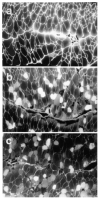
Sepsis induces skeletal muscle sarcolemmal injury. Representative micrographs illustrating the effects of sepsis on sarcolemmal integrity in the diaphragm. a, Control group. Very little tracer dye uptake within individual myofibers was found, although intense staining of extracellular connective tissue indicates good dye diffusion throughout the tissue. b, Lipopolysaccharide group. Note the presence of numerous myofibers with sarcolemmal damage, as indicated by their inability to prevent entry of the low molecular weight tracer dye. c, Cecal ligation perforation group. Myofibers with sarcolemmal damage are characterized by variable degrees of intracellular fluorescent staining, similar to the findings in the lipopolysaccharide group. Reproduced with permission from Lin et al (83).
Figure 5
Figure 5
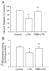
Sepsis induces alterations in skeletal muscle sarcomplasmic reticulum calcium handling. Ca2+ release and [3H]ryanodine binding were studied using sarcoplasmic reticulum membrane vesicles isolated from skeletal muscles of control and lipopolysaccharide (LPS) (7.5 mg/kg)-treated animals. Some rats were pretreated with polymyxin B (PMB), the polycationic antibiotic that neutralizes LPS before the application of LPS. A) Ryanodine (2 μM)-induced Ca2+ release from sarcoplasmic reticulum was significantly reduced in muscles from LPS-treated animals compared with controls. B) [3H]ryanodine binding was also significantly reduced in animals treated with LPS. Pretreatment with polymyxin B abated LPS-induced changes in Ca2+ release and [3H]ryanodine binding. Data are presented as mean ± standard error of the mean. *p < .05 as compared with control; **p < .05 as compared with LPS group. Reproduced with permission from Liu et al (92).
Figure 6
Figure 6
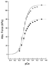
Endotoxin alters contractile protein force generation in the diaphragm. Single diaphragm fibers were isolated from control and endotoxin-treated animals, followed by permeabilization with Triton × 100 to remove the sarcolemma, sarcoplasmic reticulum, and mitochondria. Fibers were mounted on a force transducer and exposed to a series of solutions containing increasing calcium concentrations. Force per cross-sectional area was determined and expressed in kPa. The pCa is negative log of the calcium concentration (i.e., as pCa decreases, Ca2+ concentrations increase). Mean data presenting averaged absolute (Abs) force vs. pCa relationship for diaphragmatic fibers from control (◯) and endotoxemic (●) animals. Error bars represent standard error of the mean. As shown, endotoxin significantly decreased contractile force generation in single diaphragm fibers. Reproduced with permission from Supinski et al (93).
Figure 7
Figure 7
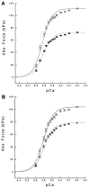
Endotoxin alters contractile protein force generation in limb muscle. Single fibers from the soleus and extensor digitorum longus were isolated from endotoxin-treated animals, permeabilized and force vs. pCa relationships determined. A) Mean data presenting averaged absolute force vs. pCa relationship for soleus fibers from control (◻) and endotoxin (■)-treated animals. B) Mean data presenting averaged absolute force vs. pCa relationship for extensor digitorum longus fibers from control (Δ) and endotoxin (▴)-treated animals. Endotoxin alters myofilament function in limb skeletal muscle. Reproduced with permission from Supinski et al (93).
Figure 8
Figure 8
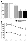
Sepsis produces decrements in oxidative phosphorylation in diaphragm mitochondria. Oxidative phosphorylation was assessed in diaphragm mitochondrial isolates from control and endotoxin-treated animals. In addition, to determine whether the defects in oxidative phosphorylation were due to changes at the level of the electron transport chain per se, the nicotinamide adenine dinucleotide (NADH) oxidase assay was also performed. A) State 3 oxygen consumption rates for diaphragm mitochondrial isolates taken from (left to right), control animals and groups of animals treated for 12, 24, 36, or 48 hrs with endotoxin. State 3 rates for 24 hrs, 36 hrs, and 48 hrs in endotoxin-treated groups were significantly lower than rates for control animals (*p < .02). Error bars indicate 1 standard error of the mean (SEM). B) Upper portion of the graph presents rates of NADH, reduced form, consumed per minute per milligram protein for diaphragm mitochondrial isolates taken from control (time zero) and (left to right) samples from animals treated with endotoxin for 12, 24, 36, or 48 hrs. The bottom portion of the graph represents state 3 oxygen consumption rates for diaphragm mitochondrial isolates taken from control and animals treated with endotoxin for 12, 24, 36, or 48 hrs (n = 4 for each group). Error bars represent 1 sem. NADH oxidase rates for groups treated with endotoxin for 36 or 48 hrs were significantly lower than rates for control animals (*p < .02 for both comparisons). The time course of reductions in NADH oxidase and state 3 respiraory rates paralleled each other, suggesting that defects in the electron transport chain per se account for most of the reduction in mitochondrial adenosine triphosphate-generating capacity. Reproduced with permission from Callahan and Supinski (99).
Figure 9
Figure 9
Endotoxin up-regulates cytokine and chemokine protein levels in skeletal muscle. Quantification of cytokine/chemokine protein levels in the diaphragm and limb muscle after lipopolysaccharide administration in vivo. Protein levels of selected proinflammatory mediators (tumor necrosis factor [TNF]-α, interleukin [IL]-6, and maximal inspiratory pressure [MIP]-2) were measured in the diaphragm (filled bars) and tibialis anterior (open bars) at 6 hrs after sham (S) or lipopolysaccharide (L) treatment. All data are group mean ± standard error of the mean (n = 6 mice/group). *p < .05 for comparisons between diaphragm and tibialis within the same condition (lipopolysaccharide or saline); p < .05 for comparisons between sham- and lipopolysaccharide-treated mice within the same type of muscle (diaphragm or tibialis). Reproduced with permission from Demoule et al (58).
Figure 10
Figure 10
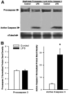
Sepsis activates caspase 3 in the diaphragm. A) Representative Western blot for procaspase 3 and active caspase 3 proteins, comparing diaphragm samples from control and endotoxin-treated (24 hrs) animals. Procaspase 3 bands were similar for samples from control and endotoxin-treated animals (top bands), but active caspase 3 protein (bottom band, top gel) was greater for samples from the endotoxin-treated group. Blotting against α-tubulin (bottom) with these samples was employed as a loading control. B) Mean densitometry data for procaspase 3 and active caspase 3 protein bands. Procaspase 3 levels were similar for samples from control and endotoxin (24 hrs)-treated animals, whereas active caspase 3 was significantly greater for samples from endotoxin-treated animals (p < .003). *Statistical difference compared with controls. LPS, lipopolysaccharide. Reproduced with permission from Supinski and Callahan (54).
Figure 11
Figure 11
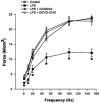
Inhibition of caspase 3 restores diaphragm-specific force generation in sepsis. Mean diaphragm force-frequency curves comparing control animals, endotoxin-treated animals (24 hrs), animals given both zVAD-fmk (a broad spectrum caspase inhibitor) and endotoxin, and animals given DEVD-CHO (a selective inhibitor of caspase 3) and endotoxin. DEVD-CHO was equivalent to zVAD-fmk in preventing endotoxin-induced reductions in diaphragm force (not significant for comparison of DEVD-CHO plus endotoxin and zVAD-fmk plus endotoxin group forces at all frequencies). *Statistical difference compared with controls. LPS, lipopolysaccharide. Reproduced with permission from Supinski and Callahan (54).
Figure 12
Figure 12
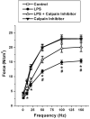
Calpain inhibition improves diaphragm-specific force generation in endotoxin-induced sepsis. Mean diaphragm force-frequency curves comparing control animals, endotoxin-treated animals (24 hrs), animals given both endotoxin and calpain inhibitor III, and animals given calpain inhibitor III alone. Force generation was significantly lower at stimulation frequencies from 10 to 150 Hz for diaphragms from lipopolysaccharide (LPS)-treated animals (filled circles) than for control animals (open squares). Diaphragms from animals given both calpain inhibitor III and lipopolysaccharide generated forces significantly higher than diaphragms from animals given endotoxin alone for frequencies from 50 to 150 Hz. Force generation for muscles taken from animals given calpain inhibitor III alone were similar to levels for control animals. *Significant statistical difference between control and endotoxin; #statistical significance between endotoxin and endotoxin plus calpain inhibitor III groups. Reproduced with permission from Supinski and Callahan (135).

References

    1. De Jonghe B, Lacherade JC, Durand MC, et al. Critical illness neuromuscular syndromes. Neurol Clin. 2008;26:507–520. - PubMed
    1. Herridge MS, Batt J, Hopkins RO. The pathophysiology of long-term neuromuscular and cognitive outcomes following critical illness. Crit Care Clin. 2008;24:179–199. - PubMed
    1. Hermans G, De Jonghe B, Bruyninckx F, et al. Clinical review: Critical illness polyneuropathy and myopathy. Crit Care. 2008;12:238. - PMC - PubMed
    1. Khan J, Harrison TB, Rich MM. Mechanisms of neuromuscular dysfunction in critical illness. Crit Care Clin. 2008;24:165–177. - PMC - PubMed
    1. Latronico N, Guarneri B. Critical illness myopathy and neuropathy. Minerva Anestesiol. 2008;74:319–323. - PubMed

Publication types