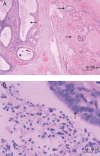
Epididymal tissue in the dilated portion of a dysgenetic kidney with an ipsilateral seminal vesicle cyst and ectopic ureteral insertion
- PMID: 20046195
- PMCID: PMC3739097
- DOI: 10.1038/aja.2009.84
Epididymal tissue in the dilated portion of a dysgenetic kidney with an ipsilateral seminal vesicle cyst and ectopic ureteral insertion
Figures


Similar articles
-
Ectopic ureter draining into seminal vesicle cyst: usefulness of MRI.Radiat Med. 1998 Jul-Aug;16(4):309-11. Radiat Med. 1998. PMID: 9814429
-
Primary mucinous adenocarcinoma of a seminal vesicle cyst associated with ectopic ureter and ipsilateral renal agenesis: a case report.Korean J Radiol. 2007 May-Jun;8(3):258-61. doi: 10.3348/kjr.2007.8.3.258. Korean J Radiol. 2007. PMID: 17554197 Free PMC article.
-
[Hydrospermatocyst with ectopic junction of the ureter and ipsilateral renal agenesis. Diagnostic difficulties and contribution of magnetic resonance imaging].J Urol (Paris). 1995;101(2):97-100. J Urol (Paris). 1995. PMID: 8522862 French.
-
[Hydrospermatocyst with ectopic insertion of the ureter associated with homolateral renal agenesis. A magnetic resonance case study].Radiol Med. 1999 Nov;98(5):427-9. Radiol Med. 1999. PMID: 10780234 Review. Italian. No abstract available.
-
[Management of High-Risk Prostate Cancer and Left Ectopic Ureter Inserting into Seminal Vesicle with Ipsilateral Hypoplastic Kidney of a Young Patient : A Case Report].Hinyokika Kiyo. 2016 Jun;62(6):329-33. Hinyokika Kiyo. 2016. PMID: 27452497 Review. Japanese.
References
-
- Shimamura M, Koizumi H, Hisazumi H. Seminal vesicle cyst associated with contralateral renal agenesis: A case report. Hinyokika Kiyo. 1984;30:1263–7. - PubMed
-
- Kosan M, Tul M, Inal G, Ugurlu O, Adsan O. A large seminal vesical cyst with contralateral renal agenesis. Int Urol Nephrol. 2006;38:591–2. - PubMed
-
- Giglio M, Medica M, Germinale F, Carmignani G. Renal dysplasia associated with ureteral ectopia and ipsilateral seminal vesicle cyst. Int J Urol. 2002;9:63–6. - PubMed
-
- Aslan DL, Pambuccian SE, Gulbahce HE, Tran ML, Manivel JC. Prostatic glands and urothelial epithelium in a seminal vesicle cyst: report of a case and review of pathologic features and prostatic ectopy. Arch Pathol Lab Med. 2006;130:194–7. - PubMed
-
- Gorrea M, Lorente R, Roel J. Seminal vesicle cyst associated with ipsilateral renal agenesis and papillary carcinoma of the bladder. Eur Radiol. 2001;11:2500–3. - PubMed
Publication types
MeSH terms
LinkOut - more resources
Full Text Sources
Medical

