

Mechanical signals as anabolic agents in bone
- PMID: 20046206
- PMCID: PMC3743048
- DOI: 10.1038/nrrheum.2009.239
Mechanical signals as anabolic agents in bone
Abstract
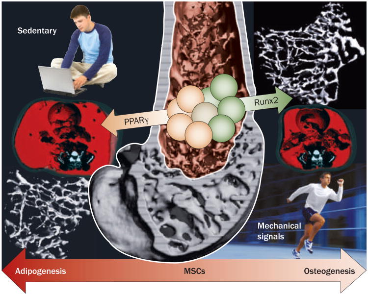
Aging and a sedentary lifestyle conspire to reduce bone quantity and quality, decrease muscle mass and strength, and undermine postural stability, culminating in an elevated risk of skeletal fracture. Concurrently, a marked reduction in the available bone-marrow-derived population of mesenchymal stem cells (MSCs) jeopardizes the regenerative potential that is critical to recovery from musculoskeletal injury and disease. A potential way to combat the deterioration involves harnessing the sensitivity of bone to mechanical signals, which is crucial in defining, maintaining and recovering bone mass. To effectively utilize mechanical signals in the clinic as a non-drug-based intervention for osteoporosis, it is essential to identify the components of the mechanical challenge that are critical to the anabolic process. Large, intense challenges to the skeleton are generally presumed to be the most osteogenic, but brief exposure to mechanical signals of high frequency and extremely low intensity, several orders of magnitude below those that arise during strenuous activity, have been shown to provide a significant anabolic stimulus to bone. Along with positively influencing osteoblast and osteocyte activity, these low-magnitude mechanical signals bias MSC differentiation towards osteoblastogenesis and away from adipogenesis. Mechanical targeting of the bone marrow stem-cell pool might, therefore, represent a novel, drug-free means of slowing the age-related decline of the musculoskeletal system.
Conflict of interest statement
Figures
Similar articles
-
microRNA-103a functions as a mechanosensitive microRNA to inhibit bone formation through targeting Runx2.J Bone Miner Res. 2015 Feb;30(2):330-45. doi: 10.1002/jbmr.2352. J Bone Miner Res. 2015. PMID: 25195535
-
Low-magnitude mechanical signals and the spine: A review of current and future applications.J Clin Neurosci. 2017 Jun;40:18-23. doi: 10.1016/j.jocn.2016.12.017. Epub 2017 Jan 12. J Clin Neurosci. 2017. PMID: 28089422 Review.
-
Low-level, high-frequency mechanical signals enhance musculoskeletal development of young women with low BMD.J Bone Miner Res. 2006 Sep;21(9):1464-74. doi: 10.1359/jbmr.060612. J Bone Miner Res. 2006. PMID: 16939405 Clinical Trial.
-
Low Intensity Vibrations Augment Mesenchymal Stem Cell Proliferation and Differentiation Capacity during in vitro Expansion.Sci Rep. 2020 Jun 10;10(1):9369. doi: 10.1038/s41598-020-66055-0. Sci Rep. 2020. PMID: 32523117 Free PMC article.
-
Regulation of mechanical signals in bone.Orthod Craniofac Res. 2009 May;12(2):94-104. doi: 10.1111/j.1601-6343.2009.01442.x. Orthod Craniofac Res. 2009. PMID: 19419452 Review.
Cited by
-
Exercise Regulation of Marrow Adipose Tissue.Front Endocrinol (Lausanne). 2016 Jul 14;7:94. doi: 10.3389/fendo.2016.00094. eCollection 2016. Front Endocrinol (Lausanne). 2016. PMID: 27471493 Free PMC article. Review.
-
In vivo assessment of the effect of controlled high- and low-frequency mechanical loading on peri-implant bone healing.J R Soc Interface. 2012 Jul 7;9(72):1697-704. doi: 10.1098/rsif.2011.0820. Epub 2012 Jan 25. J R Soc Interface. 2012. PMID: 22279157 Free PMC article.
-
Mechanical strain downregulates C/EBPβ in MSC and decreases endoplasmic reticulum stress.PLoS One. 2012;7(12):e51613. doi: 10.1371/journal.pone.0051613. Epub 2012 Dec 12. PLoS One. 2012. PMID: 23251594 Free PMC article.
-
A Novel, Direct NO Donor Regulates Osteoblast and Osteoclast Functions and Increases Bone Mass in Ovariectomized Mice.J Bone Miner Res. 2017 Jan;32(1):46-59. doi: 10.1002/jbmr.2909. Epub 2016 Sep 7. J Bone Miner Res. 2017. PMID: 27391172 Free PMC article.
-
Vibrational Force on Accelerating Orthodontic Tooth Movement: A Systematic Review and Meta-Analysis.Eur J Dent. 2023 Oct;17(4):951-963. doi: 10.1055/s-0042-1758070. Epub 2022 Dec 13. Eur J Dent. 2023. PMID: 36513343 Free PMC article.
References
-
- Kruse K, Julicher F. Oscillations in cell biology. Curr Opin Cell Biol. 2005;17:20–26. - PubMed
-
- Neel PL, Harris RW. Motion-induced inhibition of elongation and induction of dormancy in liquidambar Science. 1971;173:58–59. - PubMed
-
- Orr AW, Helmke BP, Blackman BR, Schwartz MA. Mechanisms of mechanotransduction. Dev Cell. 2006;10:11–20. - PubMed
Publication types
MeSH terms
Grants and funding
LinkOut - more resources
Full Text Sources
Other Literature Sources
