. 2009 Dec 31;50(6):818-24.
doi: 10.3349/ymj.2009.50.6.818. Epub 2009 Dec 18.

Cyclooxygenase-2 expression is related to the epithelial-to-mesenchymal transition in human colon cancers

Affiliations

Cyclooxygenase-2 expression is related to the epithelial-to-mesenchymal transition in human colon cancers

Tae Jung Jang et al. Yonsei Med J. .

Abstract

Purpose: Down-regulation of E-cadherin is a hallmark of the epithelial-to-mesenchymal transition (EMT). EMT progression in cancer cells is associated with the loss of certain epithelial markers and the acquisition of a mesenchymal phenotype, as well as migratory activities. Cyclooxygenase-2 (COX-2) expression is associated with tumor invasion and metastasis in colon cancer. This study investigated the relationship between E-cadherin and COX-2 in colon cancer cells and human colon tumors.

Materials and methods: Colon cancer cell lines and immunohistochemistry were used.

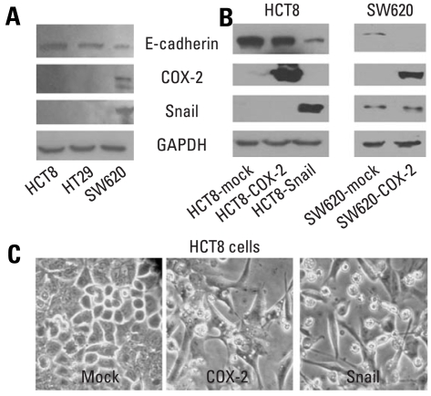
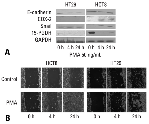
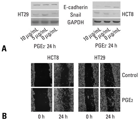
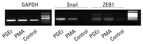
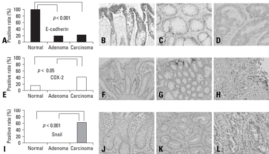
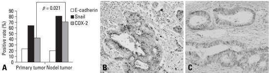
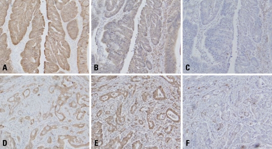
Results: E-cadherin expression was inversely related to the expressions of COX-2 and Snail in colon cancer cells. Ectopic expression of COX-2 or Snail reduced E-cadherin and induced a scattered, flattened phenotype with few intercellular contacts in colon cancer cells. Treatment of cancer cells with phorbol 12-myristate 13-acetate increased the expressions of COX-2 and Snail, decreased 15-hydroxyprostaglandin dehydrogenase expression, and increased the cells' motility. In addition, exposure to prostaglandin E(2) increased Snail expression and cell motility, and decreased E-cadherin expression. Membranous E-cadherin expression was lower in adenomas and cancers than in the adjacent, non-neoplastic epithelium. In contrast, the expressions of Snail and COX-2 were higher in cancers than in normal tissues and adenomas. The expressions of COX-2 and Snail increased in areas with abnormal E-cadherin expression. Moreover, COX-2 expression was related to higher tumor stages and was significantly higher in nodal metastatic lesions than primary cancers.

Conclusion: This study suggests that COX-2 may have a role in tumor metastasis via EMT.

Keywords: COX-2; E-cadherin; Epithelial-to-mesenchymal transition; Snail; colon cancers.


Conflict of interest statement

The authors have no financial conflicts of interest.

Figures

Fig. 1
Fig. 1
Western blot analyses of E-cadherin, COX-2 and Snail in colon cancer cells, and morphology of HCT8 transfected with cDNA for COX-2 or Snail. (A and B) Endogenous expressions of E-cadherin, COX-2, and Snail in colon cancer cells, and ectopic expressions of COX-2 and Snail in HCT8 and SW620. Forty µg of protein was separated by 10% SDS-polyacrylamide gel electrophoresis and transferred to a nitrocellulose membrane. The bottom represents GAPDH, which was used as a loading control. (C) Ectopic expression of COX-2 or Snail induced a scattered, flattened phenotype with few intercellular contacts in HCT8. COX-2, cyclooxygenase-2; SDS, sodium dodecyl sulfate; cDNA, complementary DNA; GAPDH, glyceraldehydes 3-phosphate dehydrogenase.
Fig. 2
Fig. 2
Effects of PMA on the expressions of E-cadherin, COX-2, Snail and 15-PGDH, and cell mobility in HCT8 and HT29. (A) Western blot analyses for E-cadherin, COX-2, Snail and 15-PGDH in the presence of PMA (50 ng/mL) at the indicated times. Forty µg of protein was separated by 10% SDS-polyacrylamide gel electrophoresis and transferred to a nitrocellulose membrane. The bottom represents GAPDH, which was used as a loading control. (B) Cells were incubated with serum-free medium including PMA (50 ng/mL) and allowed to migrate into the wound area for up to 24 hours at 37℃. Images were acquired immediately, and at 4 hours and 24 hours after wounding. PMA (50 ng/mL) treatment increased cell mobility in HCT8 and HT29. COX-2, cyclooxygenase-2; GAPDH, glyceraldehydes 3-phosphate dehydrogenase; PMA, phorbol 12-myristate 13-acetate; 15-PGDH, 15-hydroxyprostaglandin dehydrogenase.
Fig. 3
Fig. 3
Effects of PGE2 on the expressions of E-cadherin and Snail, and cell mobility in HCT8 and HT29. (A) Western blot analyses for E-cadherin and Snail in the presence of PGE2 at the indicated doses for 24 hours. Forty µg of protein was separated by 10% SDS-polyacrylamide gel electrophoresis and transferred to a nitrocellulose membrane. The bottom represents GAPDH, which was used as a loading control. (B) Cells were incubated with serum-free medium including with PGE2 (5 µg/mL) and allowed to migrate into the wound area for up to 24 hours at 37℃. Images were acquired immediately, and at 24 hours after wounding. PGE2 (5 µg/mL) treatment increased cell mobility in HCT8 and HT29. GAPDH, glyceraldehydes 3-phosphate dehydrogenase; PGE2, prostaglandin E2. SDS, sodium dodecyl sulfate.
Fig. 4
Fig. 4
mRNA expressions for snail and ZEB1 by RT-PCR in HT29 treated with PMA (50 ng/mL) or PGE2 (5 µg/mL for 24 hours. mRNA, messenger RNA; RT-PCR, reverse transcription-polymerase chain reaction; PMA, phorbol 12-myristate 13-acetate; PGE2, prostaglandin E2.
Fig. 5
Fig. 5
Immunohistochemical staining for E-cadherin (B, C, D), COX-2 (F, G, H) and Snail (J, K, M) in non-neoplastic adjacent mucosa (B, F, J), adenomas (C, G, K), and cancers (D, H, M). (A) Membranous E-cadherin expressions were significantly abnormal in cancers and adenomas than in non-neoplastic epithelium (p < 0.001). (E, I) In contrast, Snail and COX-2 expressions were significantly higher in cancers than in adenomas and non-neoplastic epithelium (p < 0.05, p < 0.001). COX-2, cyclooxygenase-2.
Fig. 6
Fig. 6
Immunohistochemical staining for COX-2 in nodal metastatic (B) and primary cancers (C). (A) COX-2 expression was significantly higher in nodal metastatic lesions than primary cancers (p = 0.021). COX-2, cyclooxygenase-2.
Fig. 7
Fig. 7
Topographic distributions of E-cadherin (A, D), Snail (B, E), and COX-2 (C, F) in colon cancers by immunohistochemistry. (A, B and C) Expressions of COX-2 and Snail were decreased in the areas with positive expression of E-cadherin. (D, E, F) In contrast, expressions of COX-2 and Snail increased in the areas with abnormal E-cadherin expression (D). COX-2, cyclooxygenase-2.

References

    1. Brown JR, DuBois RN. COX-2: a molecular target for colorectal cancer prevention. J Clin Oncol. 2005;23:2840–2855. - PubMed
    1. Wang D, Dubois RN. Prostaglandins and cancer. Gut. 2006;55:115–122. - PMC - PubMed
    1. Vleminckx K, Kemler R. Cadherins and tissue formation: integrating adhesion and signaling. Bioessays. 1999;21:211–220. - PubMed
    1. Thiery JP. Epithelial-mesenchymal transitions in tumour progression. Nat Rev Cancer. 2002;2:442–454. - PubMed
    1. Guarino M, Rubino B, Ballabio G. The role of epithelial-mesenchymal transition in cancer pathology. Pathology. 2007;39:305–318. - PubMed

MeSH terms