Review
. 2010 Jan-Feb;11(1):4-18.
doi: 10.3348/kjr.2010.11.1.4. Epub 2009 Dec 28.

State-of-the-art CT imaging techniques for congenital heart disease

Affiliations
Review

State-of-the-art CT imaging techniques for congenital heart disease

Hyun Woo Goo. Korean J Radiol. 2010 Jan-Feb.

Abstract

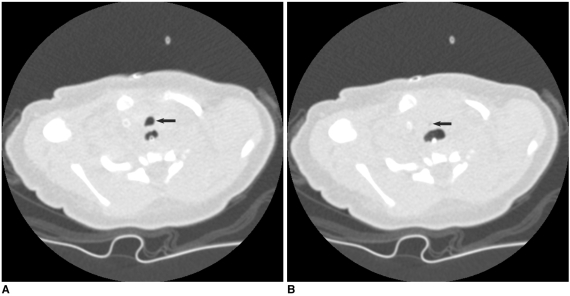
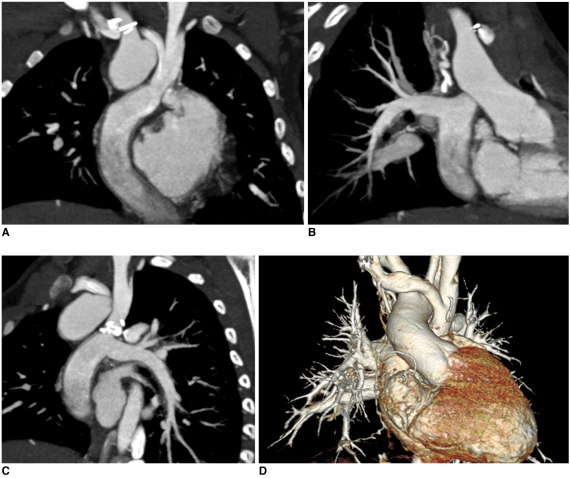
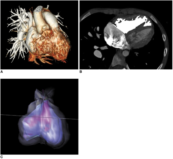
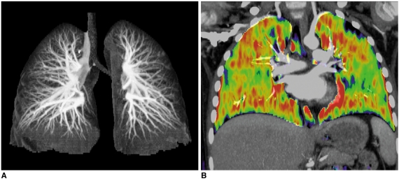
CT is increasingly being used for evaluating the cardiovascular structures and airways in the patients with congenital heart disease. Multi-slice CT has traditionally been used for the evaluation of the extracardiac vascular and airway abnormalities because of its inherent high spatial resolution and excellent air-tissue contrast. Recent developments in CT technology primarily by reducing the cardiac motion and the radiation dose usage in congenital heart disease evaluation have helped expand the indications for CT usage. Tracheobronchomalacia associated with congenital heart disease can be evaluated with cine CT. Intravenous contrast injection should be tailored to unequivocally demonstrate cardiovascular abnormalities. Knowledge of the state-of-the-art CT imaging techniques that are used for evaluating congenital heart disease is helpful not only for planning and performing CT examinations, but also for interpreting and presenting the CT image findings that consequently guide the proper medical and surgical management.

Keywords: Computed tomography (CT) techniques; Congenital heart disease; Multi-slice CT.


Figures

Fig. 1
Fig. 1
Volume-rendered CT image with non-ECG-synchronized spiral scan shows excellent anatomic details of pulmonary arteries in 12-year-old girl with repaired coarctation of aorta.
Fig. 2
Fig. 2
Effect of slice thickness on image quality of non-ECG-synchronized spiral CT in 12-year-old boy with pulmonary atresia and ventricular septal defect after Rastelli operation. A. Volume-rendered CT image reconstructed from thin, overlapped axial images with 0.6-mm slice thickness at collimation of 0.6 mm appears quite grainy. That is because slice thickness is too thin at employed CT dose and this thin slice consequently increases image noise enough to degrade image quality. There are two ways to improve image quality in this situation: one is to slightly increase slice thickness and the other is to increase radiation dose a lot. B. Slight increase in slice thickness to 0.75 mm substantially improves image quality of volume-rendered CT image. This strategy is highly recommended because its dose saving effect is great.
Fig. 3
Fig. 3
Diagrams showing retrospective ECG-gated spiral CT scan. A. As demonstrated on schema, spiral scan is acquired with low enough pitch to avoid gap in CT data. This spiral CT data is retrospectively reconstructed at specific cardiac phase by synchronizing with simultaneously acquired ECG data. B. For ECG-controlled tube current modulation, period with 100% tube current should be appropriately determined depending on heart rates. End-systolic phase (red rectangle) is used at high heart rates. Tube current can be reduced to 20% (yellow rectangle) or 4% (light green rectangle) during rest of cardiac cycle.
Fig. 4
Fig. 4
6-year-old boy with repaired tetralogy of Fallot. Sagittal (A) and short-axis (B) retrospective ECG-gated spiral CT images, which are affected by severe respiratory motion artifacts, are shown at midportion of scan range.
Fig. 5
Fig. 5
Diagrams showing prospective ECG-triggered sequential CT scan. A. Sequential scan is acquired without table movement at predefined cardiac phase, which is end-systolic phase in this diagram. Then, time period is necessary to move CT table to next scan. Thus, scan mode is commonly called 'step and shoot' mode. B. ECG shows variable heart rates ranging from 54 bpm to 111 bpm. This variability in heart rates is commonly considered to be disadvantageous for performing prospective ECG-triggered sequential scan for mid-diastolic phase. However, this is not case when end-systolic phase is target. As demonstrated on this diagram, end-systolic phases can be consistently acquired at irregular heart rate with prospective ECG-triggered sequential scan. It should be noted that absolute delay, for instance, 240 ms in this case, must be used to have this benefit.
Fig. 6
Fig. 6
Multi-phase prospective ECG-triggered sequential CT scan. A. Extended scan mode (0.38 s), in which period longer than necessary for single phase (0.2 s) is obtained, offers multi-phase study. Cardiac function can be evaluated with this scan mode at high heart rates (134 bpm in this case) because acquisition window covers both end-systole and end-diastole. B-F. After scan, five cardiac phases are retrospectively reconstructed at approximately 40-ms intervals (B, 160 ms; C, 200 ms; D, 240 ms; E, 280 ms; F 340 ms).
Fig. 7
Fig. 7
Prospective ECG-triggered sequential CT scan in free-breathing young children is relatively less susceptible to respiratory motion artifacts than is retrospectively ECG-gated spiral CT scan. With this scan mode, even side-branches (arrowheads) of coronary arteries (A) and stenosis (arrow) involving left bronchi (B) are clearly delineated without cardiac and respiratory motion artifacts in young children. Therefore, more invasive or sophisticated preparation procedures such as general anesthesia or controlled ventilation are not currently needed for only diagnostic purposes.
Fig. 8
Fig. 8
Artifacts on prospective ECG-triggered sequential CT scan. A. Various degrees of stair-step artifacts derived from respiration motion are seen on coronal CT image. Artifacts are typically much more pronounced around diaphragm. Aneurysmal dilatations involving ascending aorta (AA) and pulmonary trunk (PT) are noted in 3-year-old girl with Loeys-Dietz syndrome. B. Stair-step artifacts on prospective ECG-triggered sequential coronal CT image are remarkably decreased by applying combined ECG and respiratory triggering for same patient who underwent valve sparing aortic root replacement surgery. In contrast, difference in cardiovascular enhancement is frequently observed between adjacent slabs (arrows) due to long examination time that is further lengthened by combined triggering.
Fig. 9
Fig. 9
Combo CT scan comprised of non-ECG-synchronized spiral scan with usual scan range (A, B) and prospective ECG-triggered sequential scan with narrow scan range confined to conotruncal area of heart (C, D) in 9-months-old boy with Williams syndrome. Supravalvular aortic stenosis (arrows on C) and combined valvar (arrow on D) and subvalvar (arrowheads on D) pulmonary stenoses are clearly shown on prospective ECG-triggered sequential CT images (C, D). Dose estimates are 1.6 mSv for non-ECG-synchronized spiral scan and 0.2 mSv for prospective ECG-triggered sequential scan.
Fig. 10
Fig. 10
Cine CT (A, inspiratory phase; B, expiratory phase) for diagnosis of tracheomalacia in 16-day-old female newborn with tracheoesophageal fistula, esophageal atresia and tetralogy of Fallot. Excessive expiratory collapsibility of trachea (arrows) is displayed on cine CT images. Distended proximal esophagus is also seen posterior to trachea.
Fig. 11
Fig. 11
Dose reduction by applying low kV and ECG-controlled tube current modulation to retrospective ECG-gated spiral CT scan in 9-year-old girl with pulmonary atresia, ventricular septal defect and major aortopulmonary collateral arteries and who underwent Rastelli operation. Dose estimate of CT scan acquired with 120 kV and conventional ECG pulsing (A, B) is 9.2 mSv (heart rate: 88 bpm). On other hand, dose estimate can be reduced to 3.5 mSv by applying 100 kV and MinDose for follow-up CT (heart rate: 86 bpm). Of interest, image quality of CT images is much better and degree of cardiovascular enhancement appears much higher on follow-up CT with lower radiation dose.
Fig. 12
Fig. 12
Non-ECG-synchronized multiplanar reformatted (A-C) and volume-rendered (D) CT images clearly show patent Fontan pathway and pulmonary vessels in 5-year-old boy with functional single ventricle. Simultaneous intravenous injection of 50% diluted contrast agent through arm and leg veins was used to obtain homogeneously high enhancement of cardiovascular structures.
Fig. 13
Fig. 13
Right ventricle CT volumetry in 8-year-old boy with repaired tetralogy of Fallot. Retrospective ECG-gated spiral CT was performed because of inconsistent right ventricle volumes on serial cardiac MR examinations (end-systolic volume of right ventricle was normalized to body surface area, 86 → 128 ml/m2). A. Crisp margin of right ventricle is shown with high spatial resolution on volume-rendered CT image. B, C. Consequently, right ventricular cavity can be accurately segmented with three-dimensional region growing method. End-systolic volume of right ventricle that's normalized to body surface area calculated with CT is 86 ml/m2, which indicates that volume at second cardiac MR examination is inaccurate.
Fig. 14
Fig. 14
Dual-energy lung perfusion CT. In addition to pulmonary CT angiography (A), lung perfusion status (B) can be obtained without additional radiation dose by means of dual-energy CT technology when pulmonary thromboembolism is suspected in patients with congenital heart disease.

References

    1. Goo HW, Park IS, Ko JK, Kim YH, Seo DM, Yun TJ, et al. CT of congenital heart disease: normal anatomy and typical pathologic conditions. Radiographics. 2003;23:S147–S165. - PubMed
    1. Goo HW, Park IS, Ko JK, Kim YH, Seo DM, Park JJ. Computed tomography for the diagnosis of congenital heart disease in pediatric and adult patients. Int J Cardiovasc Imaging. 2005;21:347–365. - PubMed
    1. Leschka S, Oechslin E, Husmann L, Desbiolles L, Marincek B, Genoni M, et al. Pre- and postoperative evaluation of congenital heart disease in children and adults with 64-section CT. Radiographics. 2007;27:829–846. - PubMed
    1. Goo HW. Pediatric CT: understanding of radiation dose and optimization of imaging techniques. J Korean Radiol Soc. 2005;52:1–5. [Korean]
    1. Yang DH, Goo HW. Pediatric 16-slice CT protocol: radiation dose and image quality. J Korean Radiol Soc. 2008;59:333–347.

MeSH terms