

Incidental non-cardiac findings of a coronary angiography with a 128-slice multi-detector CT scanner: should we only concentrate on the heart?
- PMID: 20046496
- PMCID: PMC2799652
- DOI: 10.3348/kjr.2010.11.1.60
Incidental non-cardiac findings of a coronary angiography with a 128-slice multi-detector CT scanner: should we only concentrate on the heart?
Abstract
Objective: To evaluate the spectrum, prevalence, and significance of incidental non-cardiac findings (INCF) in patients referred for a non-invasive coronary angiography using a 128-slice multi-detector CT (MDCT).
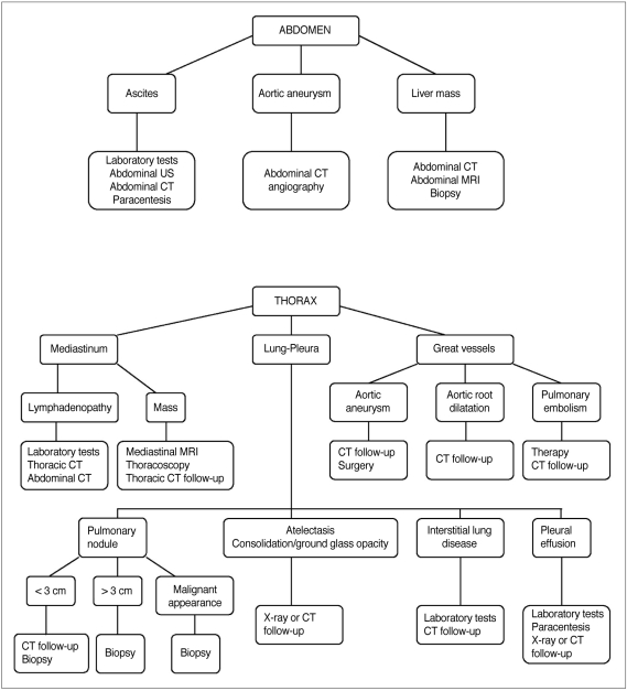
Materials and methods: The study subjects included 1,044 patients; 774 males (mean age, 59.9 years) and 270 females (mean age, 63 years), referred for a coronary CT angiography on a 128-slice MDCT scanner. The scans were acquired from the level of the carina to just below the diaphragm. To evaluate INCFs, images were reconstructed with a large field of view (> 300 mm) covering the entire thorax. Images were reviewed in the axial, coronal, and sagittal planes, using the mediastinal, lung, and bone windows. The INCFs were classified as severe, indeterminate, and mild, based on their clinical importance, and as thoracic or abdominal based on their locations.
Results: Incidental non-cardiac findings were detected in 56% of patients (588 of 1,044), including 435 males (mean age, 65.6 years) and 153 females (mean age, 67.9 years). A total of 729 INCFs were observed: 459 (63%) mild (58% thoracic, 43% abdominal), 96 (13%) indeterminate (95% thoracic, 5% abdominal), and 174 (24%) severe (87% thoracic, 13% abdominal). The prevalence of severe INCFs was 15%. Two severe INCFs were histologically verified as lung cancers.
Conclusion: The 128-slice MDCT coronary angiography, in addition to cardiac imaging, can provide important information on the pathology of the chest and upper abdomen. The presence of severe INCFs is not rare, especially in the thorax. Therefore, all organs in the scan should be thoroughly evaluated in daily clinical practice.
Keywords: Coronary computed tomography angiography; Incidental non-cardiac findings; Multi-detector computed tomography (MDCT).
Figures
Similar articles
-
Incidental Non-cardiac Findings in Coronary Computed Tomography Angiography: Is it Worth Reporting?J Clin Imaging Sci. 2019 Aug 2;9:40. doi: 10.25259/JCIS_41_2019. eCollection 2019. J Clin Imaging Sci. 2019. PMID: 31538038 Free PMC article.
-
Prevalence and significance of incidental extracardiac findings at 64-multidetector coronary CTA.J Thorac Imaging. 2007 Nov;22(4):330-4. doi: 10.1097/RTI.0b013e31813434a9. J Thorac Imaging. 2007. PMID: 18043387
-
Noncardiac findings in cardiac imaging with multidetector computed tomography.J Am Coll Cardiol. 2006 Jul 18;48(2):402-6. doi: 10.1016/j.jacc.2006.04.071. Epub 2006 May 24. J Am Coll Cardiol. 2006. PMID: 16843193
-
Thoracic CT performance and interpretation in the multi-detector era.J Thorac Imaging. 2005 Nov;20(4):253-64. doi: 10.1097/01.rti.0000174576.96498.61. J Thorac Imaging. 2005. PMID: 16282902 Review.
-
Outcomes of incidental findings on multi-detector computed tomography for transcatheter aortic valve implantation assessment: A single-centre study and review of the literature.J Med Imaging Radiat Oncol. 2019 Aug;63(4):446-453. doi: 10.1111/1754-9485.12872. Epub 2019 Mar 15. J Med Imaging Radiat Oncol. 2019. PMID: 30874377 Review.
Cited by
-
Using coronary artery calcification combined with pretest clinical risk assessment as a means of determining investigation and treatment in patients presenting with chest pain in a rural setting.Biomed Res Int. 2015;2015:582590. doi: 10.1155/2015/582590. Epub 2015 Feb 5. Biomed Res Int. 2015. PMID: 25722981 Free PMC article.
-
Unrequested findings on cardiac computed tomography: looking beyond the heart.PLoS One. 2012;7(4):e32184. doi: 10.1371/journal.pone.0032184. Epub 2012 Apr 19. PLoS One. 2012. PMID: 22536315 Free PMC article.
-
The prevalence and significance of thoracic findings in patients undergoing extended coverage computed tomography for assessment of abdominal aortic aneurysms.Br J Radiol. 2016 Jun;89(1062):20150723. doi: 10.1259/bjr.20150723. Epub 2016 Mar 18. Br J Radiol. 2016. PMID: 26987373 Free PMC article.
-
Diagnostic Accuracy of Dual-Source Computerized Tomography Coronary Angiography in Symptomatic Patients Presenting to a Referral Cardiovascular Center During Daily Clinical Practice.Iran J Radiol. 2016 Feb 29;13(2):e24350. doi: 10.5812/iranjradiol.24350. eCollection 2016 Apr. Iran J Radiol. 2016. PMID: 27679698 Free PMC article.
-
Coronavirus disease 2019 (COVID-19) pneumonia incidentally detected on coronary CT angiogram: a do-not-miss diagnosis.Emerg Radiol. 2020 Dec;27(6):721-726. doi: 10.1007/s10140-020-01802-4. Epub 2020 Jun 9. Emerg Radiol. 2020. PMID: 32519293 Free PMC article.
References
-
- Schoepf UJ, Becker CR, Ohnesorge BM, Yucel EK. CT of coronary artery disease. Radiology. 2004;232:18–37. - PubMed
-
- Achenbach S. Current and future status on cardiac computed tomography imaging for diagnosis and risk stratification. J Nucl Cardiol. 2005;12:703–713. - PubMed
-
- Wann S, Rao P, Des Prez R. Cardiac computed tomographic angiography: evaluation of non-cardiac structures. J Nucl Cardiol. 2009;16:139–150. - PubMed
-
- Kawano Y, Tamura A, Goto Y, Shinozaki K, Zaizen H, Kadota J. Incidental detection of cancers and other non-cardiac abnormalities on coronary multislice computed tomography. Am J Cardiol. 2007;99:1608–1609. - PubMed
-
- Dewey M, Schnapauff D, Teige F, Hamm B. Non-cardiac findings on coronary computed tomography and magnetic resonance imaging. Eur Radiol. 2007;17:2038–2043. - PubMed
