

Prenatal diagnosis of bilateral pulmonary agenesis: a case report
- PMID: 20046503
- PMCID: PMC2799641
- DOI: 10.3348/kjr.2010.11.1.119
Prenatal diagnosis of bilateral pulmonary agenesis: a case report
Abstract
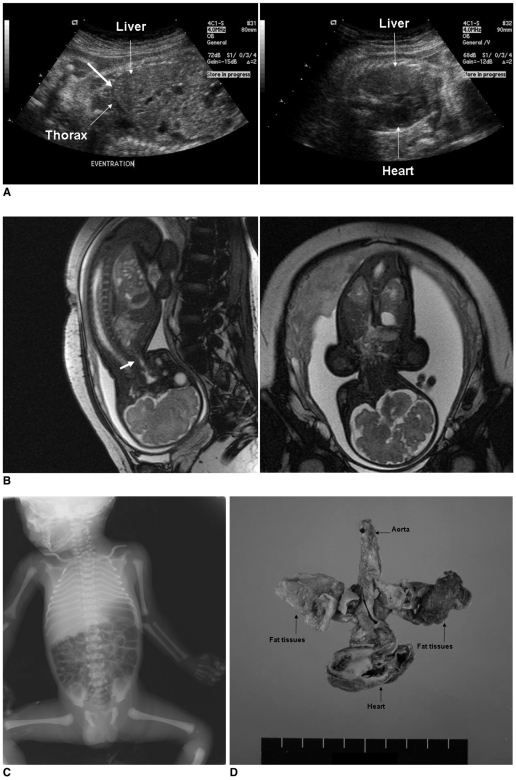
We report a case of bilateral pulmonary agenesis (BPA), which was suspected during a prenatal US examination and diagnosed by fetal magnetic resonance imaging (MRI). BPA is an extremely rare congenital anomaly and, although many fetal structural defects can be detected with a high degree of confidence after introducing high-resolution US, the prenatal diagnosis of BPA remains problematic. Other thoracic abnormalities, such as a congenital diaphragmatic hernia, congenital cystic adenomatoid malformation, and pulmonary sequestration, should be excluded from the list of possible diagnoses before coming to the conclusion of BPA, because BPA is absolutely incompatible with extrauterine life, and an accurate internal diagnosis can prevent a futile intervention from being performed.
Keywords: Bilateral pulmonary agenesis; Lung, development; Magnetic resonance (MR); Ultrasonography.
Figures
References
-
- Engellenner W, Kaplan C, Van de Vegte GL. Pulmonary agenesis association with nonimmune hydrops. Pediatr Pathol. 1989;9:725–730. - PubMed
-
- Vettraino IM, Tawil A, Comstock CH. Bilateral pulmonary agenesis: prenatal sonographic appearance simulates diaphragmatic hernia. J Ultrasound Med. 2003;22:723–726. - PubMed
-
- Nazir Z, Qazi SH, Ahmed N, Atiq M, Billoo AG. Pulmonary agenesis-vascular airway compression and gastroesophageal reflux influence outcome. J Pediatr Surg. 2006;41:1165–1169. - PubMed
-
- Lee EY, Boiselle PM, Cleveland RH. Multidetector CT evaluation of congenital lung anomalies. Radiology. 2008;247:632–648. - PubMed
-
- Laudy JA, Wladimiroff JW. The fetal lung. 1: Developmental aspects. Ultrasound Obstet Gynecol. 2000;16:284–229. - PubMed
Publication types
MeSH terms
LinkOut - more resources
Full Text Sources
Medical
