

Eosinophilic gastroenteritis with eosinophilic dermatitis
- PMID: 20046530
- PMCID: PMC2799961
- DOI: 10.3349/ymj.2010.51.1.145
Eosinophilic gastroenteritis with eosinophilic dermatitis
Abstract
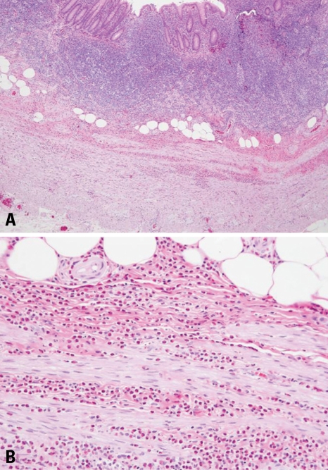
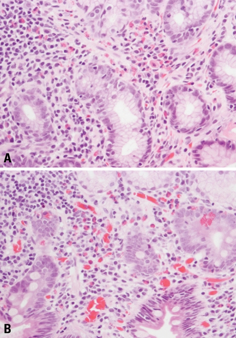
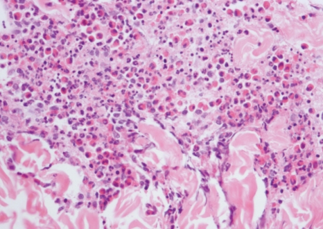
Eosinophilic gastroenteritis (EG) is characterized by eosinophilic infiltration of the bowel wall and variable gastrointestinal manifestations. Clinicians should have a high index of suspicion for EG when faced with gastrointestinal symptoms and peripheral eosinophilia to avoid incorrect diagnosis and inappropriate treatments. A 24-year-old woman was admitted to our hospital complaining of acute right lower quadrant abdominal pain and a laparoscopic appendectomy performed for a presumed diagnosis of an acute appendicitis. However, the procedure revealed bowel edema and a moderate amount of ascites without evidence of a suppurative appendicitis. Postoperatively, she showed persistent and progressive eosinophilia, exudative eosinophilic ascites, eosinophilic infiltration of the resected appendix wall, and eosinophilic infiltration of gastroduodenal mucosa. A punch biopsy of the abdominal skin also revealed inflammation with marked eosinophilic infiltration of the skin. She recovered after the treatment with a low dose of steroid for the EG with eosinophilic dermatitis. EG with eosinophilic dermatitis has not been reported yet and is considered fortuitous in this case.
Keywords: Eosinophil; ascites; corticosteroids; dermatitis; gastroenteritis.
Conflict of interest statement
The authors have no financial conflicts of interest.
Figures
References
-
- Rothenberg ME. Eosinophilic gastrointestinal disorders (EGID) J Allergy Clin Immunol. 2004;113:11–28. - PubMed
-
- Klein NC, Hargrove RL, Sleisenger MH, Jeffries GH. Eosinophlic gastroenteritis. Medicine (Baltimore) 1970;49:299–319. - PubMed
-
- Kim JD, Im EH, Lee JH, Lee TH, Kim SM, Choi YW, et al. A case of eosinophilic esophagogastroenteritis with transmural involvement. Korean J Gastrointest Endosc. 2007;35:404–409.
-
- Tran D, Salloum L, Tshibaka C, Moser R. Eosinophilic gastroenteritis mimicking acute appendicitis. Am Surg. 2000;66:990–992. - PubMed
-
- Liacouras CA. Eosinophilic esophagitis in children and adults. J Pediatr Gastroenterol Nutr. 2003;37(Suppl 1):S23–S28. - PubMed
Publication types
MeSH terms
Substances
LinkOut - more resources
Full Text Sources
Medical
