Vertebral Artery Blood flow Velocity Changes Associated with Cervical Spine rotation: A Meta-Analysis of the Evidence with implications for Professional Practice
- PMID: 20046565
- PMCID: PMC2704344
- DOI: 10.1179/106698109790818160
Vertebral Artery Blood flow Velocity Changes Associated with Cervical Spine rotation: A Meta-Analysis of the Evidence with implications for Professional Practice
Abstract
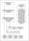
Many studies of vertebral artery (VA) blood flow changes related to cervical spine rotation have been published, but the findings are controversial and the evidence unconvincing. Recent Doppler measurements suggest that contralateral VA blood flow is compromised on full rotation in both healthy subjects and patients. More rigorous research is needed, and it was the aim of this study to conduct a meta-analysis of published data to inform professional practice. A systematic literature search, including only Doppler studies of VA blood flow velocity associated with cervical spine rotation in adults, yielded nine reports with published data. Using weighted means of the pooled data, the magnitude of the effect size (Cohen's d) was calculated for differences between patients and subjects, sitting or lying supine for testing, the parts of the VA insonated, and the changes recorded after cervical spine rotation. From this meta-analysis, VA blood flow velocity was found to be compromised more in patients than healthy individuals, on contralateral rotation, with the subject sitting, and more in the intracranial compared to the cervical part of the VA. Possible reasons for these findings are suggested, and it is advised that sustained end-of-range rotation and quick-thrust rotational manipulations be avoided until there is a stronger evidence base for clinical practice.
Keywords: Blood Flow; Cervical Spine Rotation; Physical Therapy; Vertebral Artery.
Figures


References
-
- De Kleyn A, Nieuwenhuyse AC. Vertigo and nystagmus with various head positions. Acta Otolaryngol. 1927;11:155–157.
-
- Tissington-Tatlow WF, Brammer HG. Syndrome of vertebral artery compression. Neurology. 1957;7:331–340. - PubMed
-
- Toole JF, Tucker SH. Influence of head position upon cerebral circulation. Arch Neurol. 1960;2:616–623. - PubMed
-
- Brown BSTJ, Tissington-Tatlow WF. Radiographic studies of the vertebral arteries in cadavers. Radiology. 1963;81:80–88. - PubMed
-
- Selecki BR. The effects of rotation of the atlas on the axis: Experimental work. Med J Aust. 1969;1:1012–1015. - PubMed
LinkOut - more resources
Full Text Sources
