

Xenotransplantation of cryopreserved composite organs on the rabbit
- PMID: 20046675
- PMCID: PMC2781092
- DOI: 10.4161/org.5.3.9584
Xenotransplantation of cryopreserved composite organs on the rabbit
Abstract
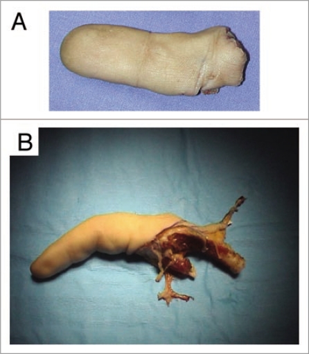
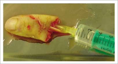
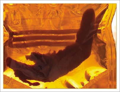
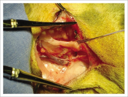
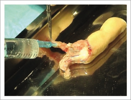
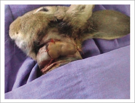
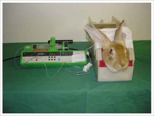
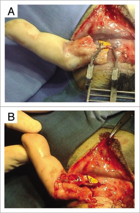
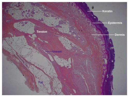
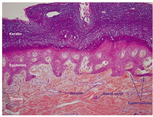
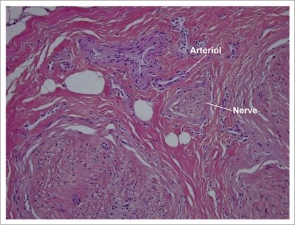
Nowadays, It is easy to define optimal conditions (cryoprotective agent, speed and steps of freezing, speed of warming) for the cryopreservation of a homogeneous cell population or a one cell-layer tissue. Meanwhile, It is still hard to obtain cryopreservation of composite organs. Each tissue has its own requirements and its own reactivity to the cryopreservation process. The challenge consists of, on the one hand, to select the ideal combination of cryoprotective agents that can fit the needs of the different tissues, and the definition of adequate technical parameters, on the other hand. All the experimental trials have studied the survival rate of non-vascularized cryopreserved tissues. The aim of our experimental work is to demonstrate the feasibility of cryopreserving a composite organ with its nutrient vessels "artery and veins" in order after thawing to revitalize it by reestablishing the blood irrigation by microsurgical vascular anastomosis. We report our experimental results on the cryopreservation of composite organs-amputated digits-xenotransplanted in the rabbit. Digital segments were cryopreserved, then revitalized after warming using vascular microsurgical techniques. Preliminary results are encouraging and may pave the way in the future to the microvascular allotransplantation of cryopreserved composite organs.
Keywords: animals; composite tissues; cryopreservation; digits; microsurgery; organs; skin; vascular anastomoses; vessels; xenotransplantation.
Figures
References
-
- Oufquir A, Bakhach J, Panconi B, Guimberteau JC, Baudet J. Sauvetage des revascularisations digitales par administration intra-artérielle de fibrinolytiques. Ann Chir Plast Esthet. 2006:51471–51481. (Fre). - PubMed
-
- Furnas H, Rosen J. Monitoring in microvascular surgery. Ann Plast Surg. 1991;26:265–272. - PubMed
-
- Yii Ni W, Evans GRD, Miller MJ, Reece GP, Langstein H, Chang D, et al. Thrombolytic therapy: what is its role in free flap salvage? Ann Plast Surg. 2001;46:601–604. - PubMed
-
- Serletti JM, Moran SL, Orlando GS, O'Connor T, Herrera R. Urokinase protocol for free-flap salvage following prolonged venous thrombosis. Plast Reconstr Surg. 1998;102:1947–1953. - PubMed
-
- Zhang F, Attkiss KJ, Walker M, Buncke HJ. Effect of cryopreservation on survival of composite tissue grafts. J Reconstr Microsurg. 1998;14:559–564. - PubMed
LinkOut - more resources
Full Text Sources