

Effects of an intravitreal bevacizumab injection combined with panretinal photocoagulation on high-risk proliferative diabetic retinopathy
- PMID: 20046686
- PMCID: PMC2789950
- DOI: 10.3341/kjo.2009.23.4.266
Effects of an intravitreal bevacizumab injection combined with panretinal photocoagulation on high-risk proliferative diabetic retinopathy
Abstract
Purpose: To investigate the short-term effects of panretinal photocoagulation (PRP) combined with an intravitreal injection of Avastin(bevacizumab) as an adjuvant to high-risk proliferative diabetic retinopathy (PDR).
Methods: The data was collected retrospectively from the eyes of high-risk PDR patients, which were divided into two groups. One eye was treated with only PRP (PRP only group) and the fellow eye of same patient was treated with both PRP and intravitreal bevacizumab injection (Adjuvant group). Best corrected visual acuity (BCVA), IOP (intraocular pressure), and new vessel (NV) size in fluorescein angiography were recorded immediately and at the six-week follow-up visit. Adverse events associated with intravitreal injection were investigated.
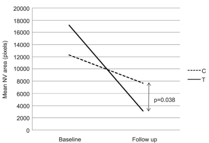
Results: Of 12 patients with high-risk PDR, five were male and seven were female. There were no statistically significant BCVA or IOP changes after treatment in either group (p=0.916, 0.888). The reduction of NV size was found in both groups, but NV size in the adjuvant group showed a greater decrease than that of the PRP only group (p=0.038). Three patients had adverse events after intravitreal injection. Two patients had mild anterior uveitis and one patient had a serious complication of branched retinal artery obstruction (BRAO).
Conclusions: Intravitreal bevacizumab injection with PRP resulted in marked regression of neovascularization compared with PRP alone. One serious side effect, BRAO, was noted in this study. Further studies are needed to determine the effect of repeated intravitreal bevacizumab injections and the proper number of bevacizumab injections as an adjuvant.
Keywords: Bevacizumab; Neovascularization; Panretinal photocoagulation; Proliferative diabetic retinopathy.
Figures
References
-
- Kaiser RS, Maguire MG, Grunwald JE, et al. One-year outcomes after panretinal photocoagulation in proliferative diabetic retinopathy. Am J Ophthalmol. 2000;129:178–185. - PubMed
-
- Early photocoagulation for diabetic retinopathy: ETDRS report number 9. Early Treatment Diabetic Retinopathy Study Research Group. Ophthalmology. 1991;98(5 Suppl):766–785. - PubMed
-
- Kleiner RC, Elman MJ, Murphy RP, Ferris FL., 3rd Transient severe visual loss after panretinal photocoagulation. Am J Ophthalmol. 1988;106:298–306. - PubMed
-
- McDonald HR, Schatz H. Macular edema following panretinal photocoagulation. Retina. 1985;5:5–10. - PubMed
-
- Flynn HW, Jr, Chew EY, Simons BD, et al. Pars plana vitrectomy in the Early Treatment Diabetic Retinopathy Study. EDTRS report number 17. The Early Treatment Diabetic Retinopathy Study Research Group. Ophthalmology. 1992;99:1351–1357. - PubMed
Publication types
MeSH terms
Substances
LinkOut - more resources
Full Text Sources
Medical
Research Materials
