

A case of pseudo-Duane's retraction syndrome with old medial orbital wall fracture
- PMID: 20046701
- PMCID: PMC2789965
- DOI: 10.3341/kjo.2009.23.4.329
A case of pseudo-Duane's retraction syndrome with old medial orbital wall fracture
Abstract
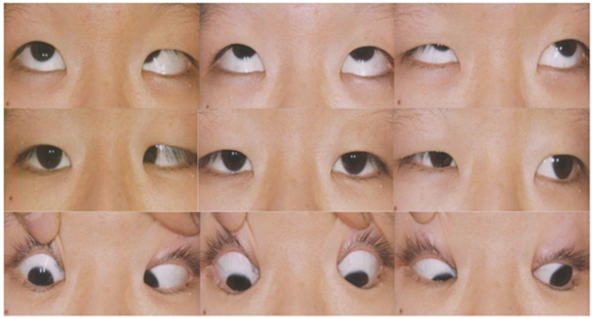
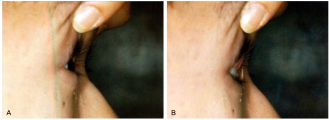
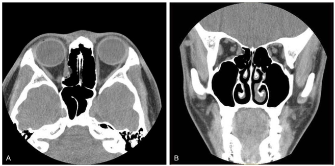
We report a case of pseudo-Duane's retraction syndrome with entrapment of the medial rectus muscle in an old medial orbital wall fracture presenting identical clinical symptoms as Duane's retraction syndrome. A 15-year-old boy presented with persistent limited right eye movement since a young age. Examination showed marked limited abduction, mildly limited adduction, and globe retraction accompanied by narrowing of the palpebral fissure during attempted adduction in the right eye. He showed a right esotropia of 16 prism diopters and his head turned slightly to the right. A slight enophthalmos was noted in his right eye. A computed tomography scan demonstrated entrapment of the medial rectus muscle and surrounding tissues in an old medial orbital wall fracture. A forced duction test revealed a marked restriction of abduction in the right eye. A 5 mm recession of the right medial rectus muscle was performed. Postoperatively, the patient's head turn and esotropia in the primary position were successfully corrected, but there was still some limitations to his ocular movement. The importance of several tests such as the forced duction test and an imaging study should be emphasized in making a diagnosis for limitation of eye movement.
Keywords: Duane's retraction syndrome; Medial orbital wall fracture; Pseudo-Duane's retraction syndrome.
Figures
References
-
- DeRespinis PA, Caputo AR, Wagner RS, Guo S. Duane's retraction syndrome. Surv Ophthalmol. 1993;38:257–288. - PubMed
-
- Gittinger JW, Jr, Hughes JP, Suran EL. Medial orbital wall blow-out fracture producing an acquired retraction syndrome. J Clin Neuroophthalmol. 1986;6:153–156. - PubMed
-
- Chatterjee PK, Bhunia J, Bhattacharyya I. Bilateral inverse Duane's retraction syndrome: a case report. Indian J Ophthalmol. 1991;39:183–185. - PubMed
Publication types
MeSH terms
LinkOut - more resources
Full Text Sources
