

Functional and structural changes in the chinchilla cochlea and vestibular system following round window application of carboplatin
- PMID: 20046821
- PMCID: PMC2800309
- DOI: 10.3109/16513860903335795
Functional and structural changes in the chinchilla cochlea and vestibular system following round window application of carboplatin
Abstract
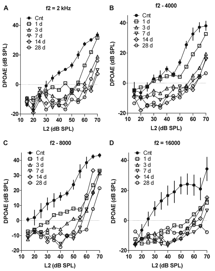
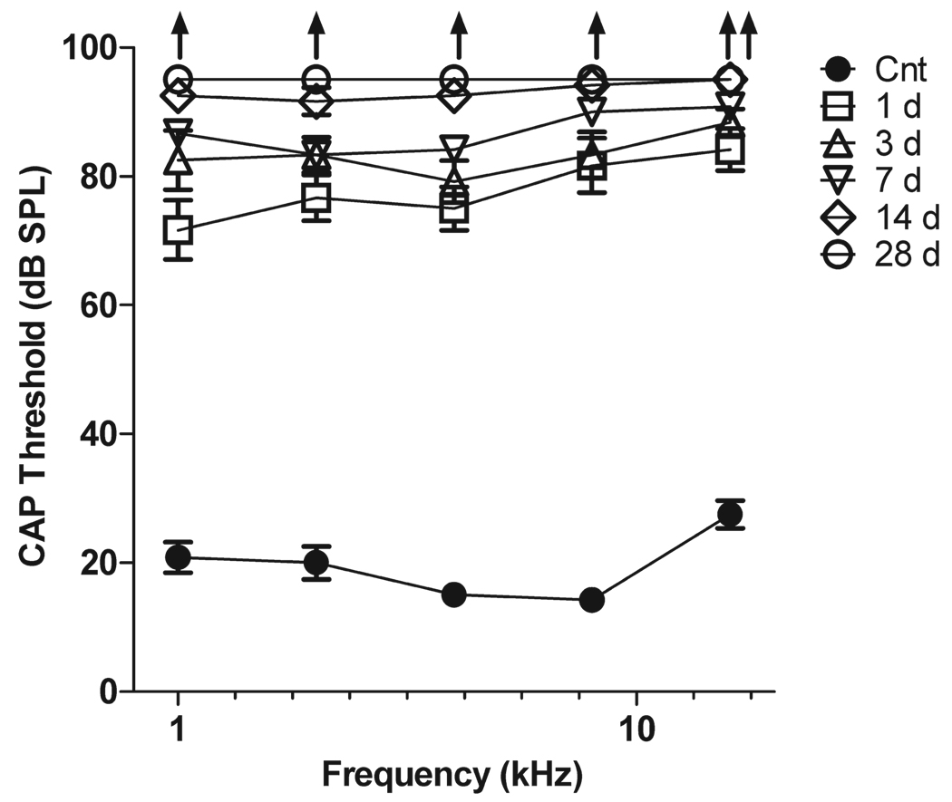
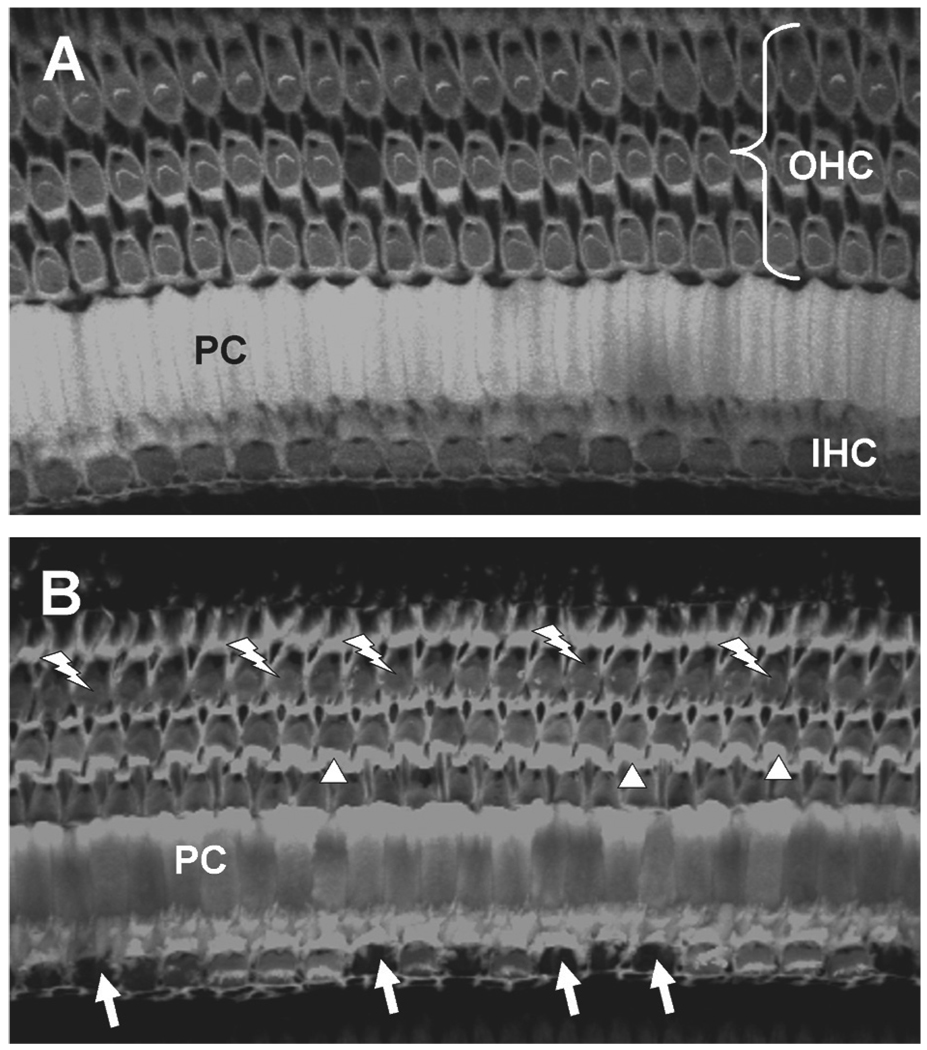
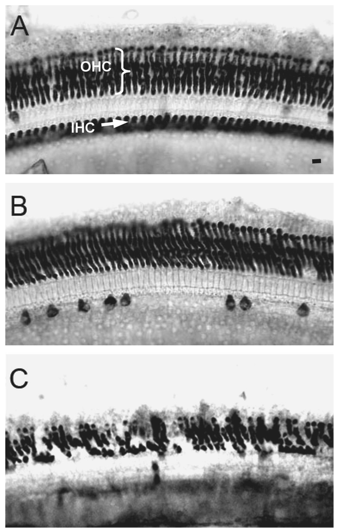
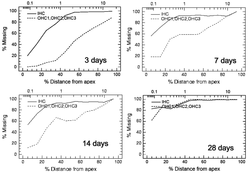
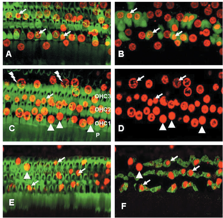
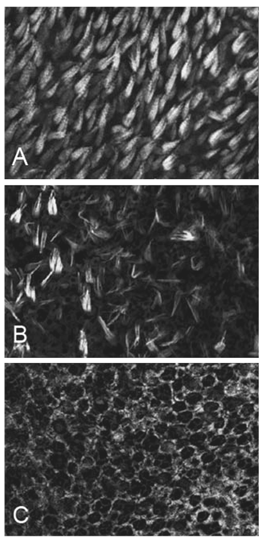
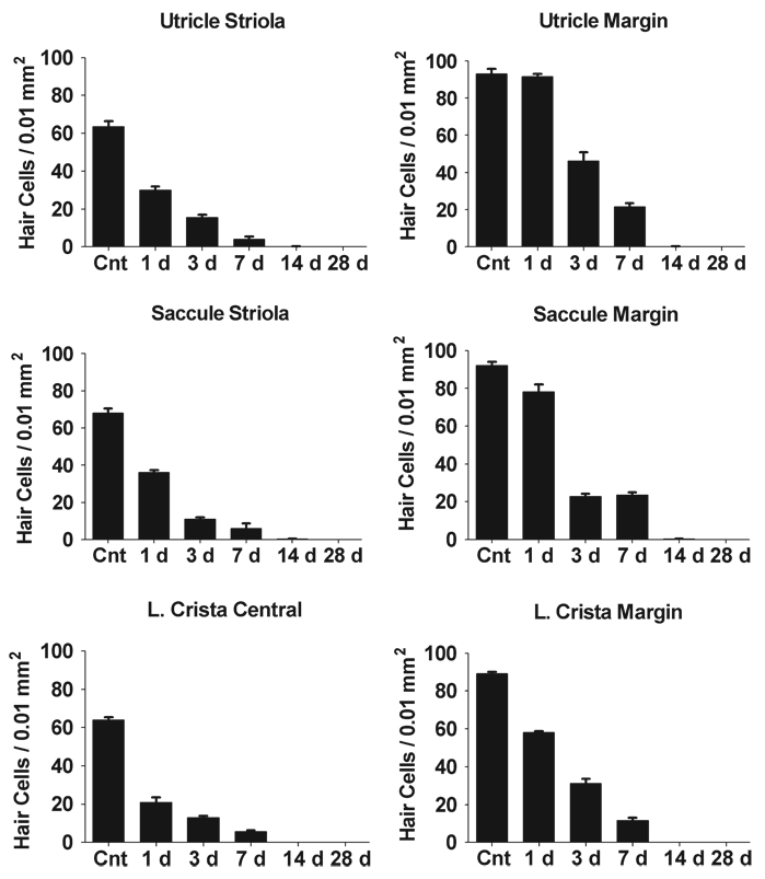
OBJECTIVE: In chinchillas, moderate doses of carboplatin administered systemically selectively destroy inner hair cells and type I vestibular hair cells; however, it is unclear whether this unique damage pattern persists if carboplatin is applied directly to the cochlea, how quickly the damage develops and what cell death pathways are involved. STUDY DESIGN: To address these questions, carboplatin (5 mg/ml, 50 µl) was applied to the round window. RESULTS: Carboplatin caused a rapid decline in distortion product otoacoustic emissions, significantly increased compound action potential thresholds and caused massive inner hair cell loss and less severe outer hair cell loss. Hair cell loss was initially more severe in the base than the apex of the cochlea, but by 28 days post-treatment most cochlear hair cells were missing and hair cell density in the utricle, saccule and lateral crista was greatly reduced. At one day post-treatment, many hair cell nuclei were condensed or fragmented indicative of apoptosis, and expressed initiator caspase-8 and executioner caspase-3, but not initiator caspase-9. Carboplatin-treated animals circled towards the treated ear and during the swim test rolled towards the treated ear. CONCLUSION: These results indicate that local application of carboplatin causes loss of hair cells that begins near the base of the cochlea and spreads towards the apex with increasing survival time. Hair cell loss is initiated by caspase-8 followed by executioner caspase-3.
Conflict of interest statement
Figures
Similar articles
-
[Inner hair cells loss by carboplatin and the changes of cochlear compound action potential in chinchillas].Zhonghua Er Bi Yan Hou Tou Jing Wai Ke Za Zhi. 2020 May 7;55(5):506-513. doi: 10.3760/cma.j.cn.115330-20200426-00332. Zhonghua Er Bi Yan Hou Tou Jing Wai Ke Za Zhi. 2020. PMID: 32842367 Chinese.
-
OTOTOXIC EFFECTS OF CARBOPLATIN IN ORGANOTYPIC CULTURES IN CHINCHILLAS AND RATS.J Otol. 2012 Dec;7(2):92-101. doi: 10.1016/S1672-2930(12)50023-1. J Otol. 2012. PMID: 25593588 Free PMC article.
-
Quantitative relationship of carboplatin dose to magnitude of inner and outer hair cell loss and the reduction in distortion product otoacoustic emission amplitude in chinchillas.Hear Res. 1997 Oct;112(1-2):199-215. doi: 10.1016/s0378-5955(97)00123-8. Hear Res. 1997. PMID: 9367242
-
Selective loss of inner hair cells and type-I ganglion neurons in carboplatin-treated chinchillas. Mechanisms of damage and protection.Ann N Y Acad Sci. 1999 Nov 28;884:152-70. doi: 10.1111/j.1749-6632.1999.tb08640.x. Ann N Y Acad Sci. 1999. PMID: 10842592 Review.
-
Review: ototoxic characteristics of platinum antitumor drugs.Anat Rec (Hoboken). 2012 Nov;295(11):1851-67. doi: 10.1002/ar.22577. Epub 2012 Oct 8. Anat Rec (Hoboken). 2012. PMID: 23044998 Review.
Cited by
-
Ototoxic destruction by co-administration of kanamycin and ethacrynic acid in rats.J Zhejiang Univ Sci B. 2011 Oct;12(10):853-61. doi: 10.1631/jzus.B1100040. J Zhejiang Univ Sci B. 2011. PMID: 21960349 Free PMC article.
-
Mefloquine damage vestibular hair cells in organotypic cultures.Neurotox Res. 2011 Jul;20(1):51-8. doi: 10.1007/s12640-010-9221-z. Epub 2010 Sep 22. Neurotox Res. 2011. PMID: 20859773 Free PMC article.
-
Up-regulation of GAP-43 in the chinchilla ventral cochlear nucleus after carboplatin-induced hearing loss: correlations with inner hair cell loss and outer hair cell loss.Hear Res. 2013 Aug;302:74-82. doi: 10.1016/j.heares.2013.05.002. Epub 2013 May 23. Hear Res. 2013. PMID: 23707995 Free PMC article.
-
Species Differences in the Organization of the Ventral Cochlear Nucleus.Anat Rec (Hoboken). 2018 May;301(5):862-886. doi: 10.1002/ar.23751. Epub 2018 Jan 6. Anat Rec (Hoboken). 2018. PMID: 29236365 Free PMC article.
-
Blocking caspase-3-dependent pathway preserves hair cells from salicylate-induced apoptosis in the guinea pig cochlea.Mol Cell Biochem. 2011 Jul;353(1-2):291-303. doi: 10.1007/s11010-011-0798-1. Epub 2011 Apr 19. Mol Cell Biochem. 2011. PMID: 21503676
References
-
- Macdonald MR, Harrison RV, Wake M, Bliss B, Macdonald RE. Ototoxicity of carboplatin: comparing animal and clinical models at the Hospital for Sick Children. J Otolaryngol. 1994;23:151–159. - PubMed
-
- Taudy M, Syka J, Popelar J, Ulehlova L. Carboplatin and cisplatin oto-toxicity in guinea pigs. Audiology. 1992;31:293–299. - PubMed
-
- Mount RJ, Takeno S, Wake M, Harrison RV. Carboplatin ototoxicity in the chinchilla: lesions of the vestibular sensory epithelium. Acta Otolaryngol Suppl. 1995;519:60–65. - PubMed
-
- Schweitzer VG, Rarey KE, Dolan DF, Abrams G, Litterst CJ, Sheridan C. Ototoxicity of cisplatin versus platinum analogs CBDCA (JM-8) and CHIP (JM-9) Otolaryngol Head Neck Surg. 1986;94:458–470. - PubMed
Grants and funding
LinkOut - more resources
Full Text Sources
Research Materials
Miscellaneous