. 2009 Dec 31;4(12):e8543.
doi: 10.1371/journal.pone.0008543.

Abnormal placental development and early embryonic lethality in EpCAM-null mice

Affiliations

Abnormal placental development and early embryonic lethality in EpCAM-null mice

Keisuke Nagao et al. PLoS One. .

Abstract

Background: EpCAM (CD326) is encoded by the tacstd1 gene and expressed by a variety of normal and malignant epithelial cells and some leukocytes. Results of previous in vitro experiments suggested that EpCAM is an intercellular adhesion molecule. EpCAM has been extensively studied as a potential tumor marker and immunotherapy target, and more recent studies suggest that EpCAM expression may be characteristic of cancer stem cells.

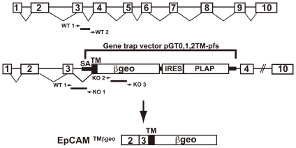
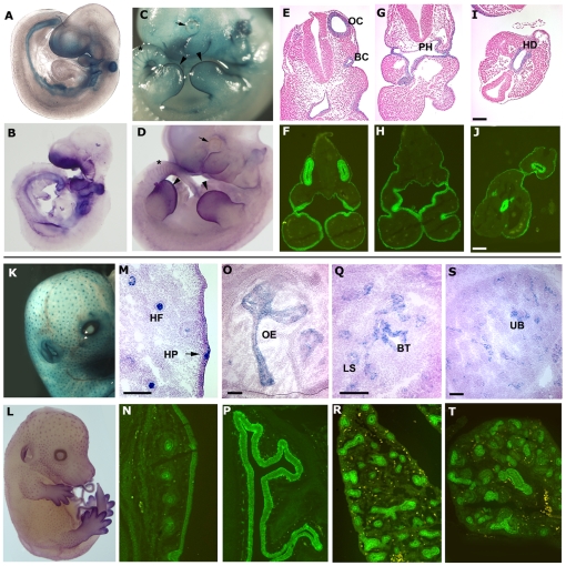
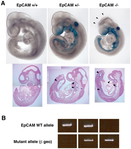
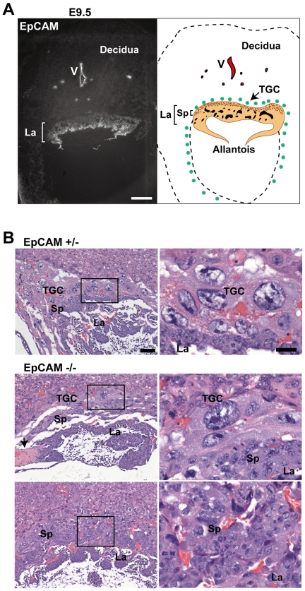
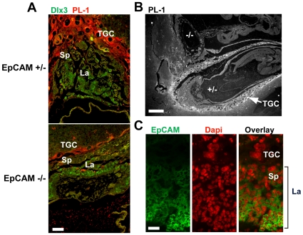
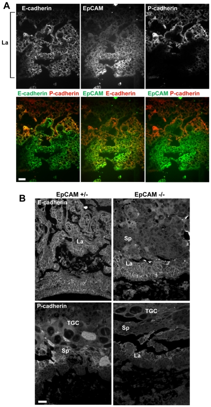
Methodology/principal findings: To gain insights into EpCAM function in vivo, we generated EpCAM -/- mice utilizing an embryonic stem cell line with a tacstd1 allele that had been disrupted. Gene trapping resulted in a protein comprised of the N-terminus of EpCAM encoded by 2 exons of the tacstd1 gene fused in frame to betageo. EpCAM +/- mice were viable and fertile and exhibited no obvious abnormalities. Examination of EpCAM +/- embryos revealed that betageo was expressed in several epithelial structures including developing ears (otocysts), eyes, branchial arches, gut, apical ectodermal ridges, lungs, pancreas, hair follicles and others. All EpCAM -/- mice died in utero by E12.5, and were small, developmentally delayed, and displayed prominent placental abnormalities. In developing placentas, EpCAM was expressed throughout the labyrinthine layer and by spongiotrophoblasts as well. Placentas of EpCAM -/- embryos were compact, with thin labyrinthine layers lacking prominent vascularity. Parietal trophoblast giant cells were also dramatically reduced in EpCAM -/- placentas.

Conclusion: EpCAM was required for differentiation or survival of parietal trophoblast giant cells, normal development of the placental labyrinth and establishment of a competent maternal-fetal circulation. The findings in EpCAM-reporter mice suggest involvement of this molecule in development of vital organs including the gut, kidneys, pancreas, lungs, eyes, and limbs.


Conflict of interest statement

Competing Interests: The authors have declared that no competing interests exist.

Figures

Figure 1
Figure 1. Disruption of the tacstd1gene that encodes murine EpCAM.
The schematic indicates the site of insertion of the gene trapping vector as well as the structure of the mRNA that encodes the EpCAM/βgeo fusion protein. Locations of primers used for genotyping are also depicted.
Figure 2
Figure 2. Expression of EpCAM during embryonic development.
(A–D) Whole mount LacZ staining (A&C, in blue) and in situ hybridization (B&D, in purple) of E9.5 (A&B) and E11.5 (C&D) embryos. EpCAM expression in the apical ectodermal ridges (arrowheads), around the eyes (arrows) and inter-somitic regions (asterisks) is highlighted in C and D. Corresponding transverse sections with LacZ and light H&E staining (E, G, I), and immunofluorescence staining using rat anti-mouse EpCAM mAb (F, H, J, in green). OC = otocyst, BC = 1st branchial cleft, Ph = pharynx, HD = hindgut diverticulum. (K–T) EpCAM expression in epithelia in a variety of organs at E14.5. K, M, O, Q, and S show LacZ and N, P, R, T show immunofluorescence staining. Primordial hair follicles are prominently stained in the E14.5 embryo as determined by LacZ staining (K&M), in situ hybridization (L), and immunofluorescence (N). M to T shows EpCAM expression in skin (M&N), nasal plexus (O&P), lungs (Q&R), and kidneys (S&T). Bars = 100 µm. HF = hair follicle, HP = hair placode, OE = olfactory epithelium, BT = bronchiolar tubules, LS = lung saccules, UB = ureteric bud branches.
Figure 3
Figure 3. Developmental delay in EpCAM-deficient embryos.
(A) Embryonic development in wild type, haplosufficient and EpCAM-deficient littermate embryos (E9.5). Arrows indicate open neural tube. Sagittal sections show EpCAM (βgeo) expression in gut endoderm in EpCAM +/− and EpCAM −/− embryos (arrowheads). The embryos depicted were littermates and photos of whole mounts were taken at identical magnifications. (B) Genotypes of embryos depicted in (A).
Figure 4
Figure 4. Defective placental development in the absence of EpCAM.
(A) EpCAM expression in wild type C57BL/6 placentas at E 9.5 as shown by immunofluorescence staining (left). Bar = 200 µm. Right panel shows a schematic of E9.5 placenta. La = labyrinth, Sp = spongiotrophoblasts, TGC = parietal trophoblast giant cells, V = vasculature. (B) Morphology in H&E stained sections of EpCAM +/− and −/− placentas. Bars = 50 and 20 µm. Panels on the right represent high power view of insets in the left panels. Arrow in B highlights intravascular microthrombus.
Figure 5
Figure 5. Diminished parietal trophoblast giant cell population in EpCAM −/− placentas.
(A) Sections of E9.5 placenta double-stained with anti-Dlx3 antibody and anti-PL-1 antibody after antigen retrieval. (B) Anti-PL-1 staining of a section that contained two embryos (EpCAM −/−, upper embryo; EpCAM +/−, lower embryo) in one E9.5 uterine decidual swelling. (C) EpCAM and DAPI staining of a frozen section of an E9.5 wild type placenta. Bars = 50 (A), 200 (B), and 20 µm (C).
Figure 6
Figure 6. Cadherin expression in EpCAM-sufficient and deficient placentas.
(A) Frozen sections from E9.5 wild type placentas stained for EpCAM, E-, and P-cadherins (La = labyrinth). (B) Sections from formalin-fixed, paraffin-embedded tissues stained for E-cadherin and P-cadherin after antigen retrieval. Bars = 20 µm.

Similar articles

Cited by

References

    1. Edwards DP, Grzyb KT, Dressler LG, Mansel RE, Zava DT, et al. Monoclonal antibody identification and characterization of a Mr 43,000 membrane glycoprotein associated with human breast cancer. Cancer Res. 1986;46:1306–1317. - PubMed
    1. Herlyn M, Steplewski Z, Herlyn D, Koprowski H. Colorectal carcinoma-specific antigen: detection by means of monoclonal antibodies. Proc Natl Acad Sci U S A. 1979;76:1438–1442. - PMC - PubMed
    1. Ross AH, Herlyn D, Iliopoulos D, Koprowski H. Isolation and characterization of a carcinoma-associated antigen. Biochem Biophys Res Commun. 1986;135:297–303. - PubMed
    1. Bergsagel PL, Victor-Kobrin C, Timblin CR, Trepel J, Kuehl WM. A murine cDNA encodes a pan-epithelial glycoprotein that is also expressed on plasma cells. J Immunol. 1992;148:590–596. - PubMed
    1. Borkowski TA, Nelson AJ, Farr AG, Udey MC. Expression of gp40, the murine homologue of human epithelial cell adhesion molecule (Ep-CAM), by murine dendritic cells. Eur J Immunol. 1996;26:110–114. - PubMed

Publication types

MeSH terms