. 2009 Dec 31;4(12):e8531.
doi: 10.1371/journal.pone.0008531.

Mechanisms of c-myc degradation by nickel compounds and hypoxia

Affiliations

Mechanisms of c-myc degradation by nickel compounds and hypoxia

Qin Li et al. PLoS One. .

Abstract

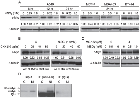
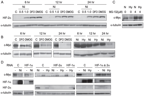
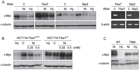
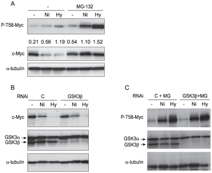
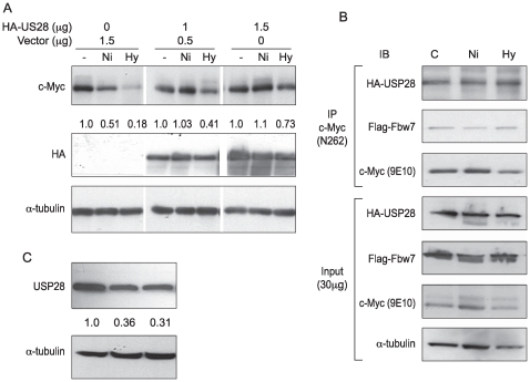
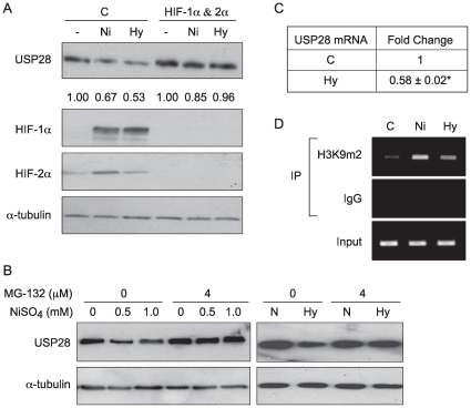
Nickel (Ni) compounds have been found to cause cancer in humans and animal models and to transform cells in culture. At least part of this effect is mediated by stabilization of hypoxia inducible factor (HIF1a) and activating its downstream signaling. Recent studies reported that hypoxia signaling might either antagonize or enhance c-myc activity depending on cell context. We investigated the effect of nickel on c-myc levels, and demonstrated that nickel, hypoxia, and other hypoxia mimetics degraded c-myc protein in a number of cancer cells (A549, MCF-7, MDA-453, and BT-474). The degradation of the c-Myc protein was mediated by the 26S proteosome. Interestingly, knockdown of both HIF-1alpha and HIF-2alpha attenuated c-Myc degradation induced by Nickel and hypoxia, suggesting the functional HIF-1alpha and HIF-2alpha was required for c-myc degradation. Further studies revealed two potential pathways mediated nickel and hypoxia induced c-myc degradation. Phosphorylation of c-myc at T58 was significantly increased in cells exposed to nickel or hypoxia, leading to increased ubiquitination through Fbw7 ubiquitin ligase. In addition, nickel and hypoxia exposure decreased USP28, a c-myc de-ubiquitinating enzyme, contributing to a higher steady state level of c-myc ubiquitination and promoting c-myc degradation. Furthermore, the reduction of USP28 protein by hypoxia signaling is due to both protein degradation and transcriptional repression. Nickel and hypoxia exposure significantly increased the levels of dimethylated H3 lysine 9 at the USP28 promoter and repressed its expression. Our study demonstrated that Nickel and hypoxia exposure increased c-myc T58 phosphorylation and decreased USP28 protein levels in cancer cells, which both lead to enhanced c-myc ubiquitination and proteasomal degradation.


Conflict of interest statement

Competing Interests: The authors have declared that no competing interests exist.

Figures

Figure 1
Figure 1. The decrease in c-Myc protein levels in A549 cells was primarily due to proteasomal degradation.
(A) Various cancerous cell lines were treated with NiSO4 at indicated concentrations for 6, 12 or 24 hr. After treatment, the cells were lysed and whole cell lysates were analyzed by Western blot using anti-c-Myc antibody. The same membranes were then stripped and re-probed with α-tubulin antibody to assess protein loading. The densitometric data, normalized to α-tubulin level, is expressed as fold-change from control; these values are presented between the blots. (B) A549 cells were treated with 1.0 mM NiSO4 for 24 hr and then cycloheximide (CHX) was added to a final concentration of 10 µg/ml. Cells were lysed at selected time intervals, whole cell lysate was then isolated for Western blot. The relative intensity of the bands was quantitated using Image J software and normalized to the loading control. The numbers quantitating the intensity of the bands were given below the corresponding bands and were used to fit the exponential lines by Excel, and half-lives were calculated and shown below the figure. (C) A549 cells were pretreated with 4 µM of MG-132 for 2 hr and then NiSO4 was added into the medium for another 24 hr. After treatment, cells were lysed and 50 µg of whole cell lysate was analyzed by Western blotting. (D) Ni compounds increased ubiquitinated c-Myc (Ub-c-Myc) in A549 cells. A549 cells were transfected with c-Myc plasmid. 24 hr after transfection, cells were treated with NiSO4 (1 mM) in the presence of MG-132 (4 µM) for another 24 hr. Cells were then lysed and immunoprecipitated with anti-Ubiquitin (anti-Ub) antibody or non-specific IgG. Precipitates were analyzed by immunoblotting with anti-c-Myc (c-33) antibody (mouse).
Figure 2
Figure 2. Knocking down both HIF-1α and HIF-2α attenuated c-Myc degradation by Ni ions and hypoxia in A549 cells.
(A) NiSO4 (0.5 and 1 mM), desferrioxamine (DFO: 100 µM), and dimethyloxallyl glycine (DMOG: 1 mM) induced HIF-2α in A549 cells in a time-dependent manner. (B) 100 µM DFO, 1 mM DMOG, and hypoxia (1% O2) decreased c-Myc protein levels in A549 cells in a time-dependent manner. N is normoxia, while Hy is hypoxia. (C) Proteasome inhibitor MG-132 prevented c-Myc degradation by hypoxia in A549 cells. (D) A549 cells were transfected with non-coding control siRNA or siRNA targeting human HIF-1α, HIF-2α or mixture (HIF-1α RNAi and HIF-2α RNAi together) for 48 hr, and then were treated with 1.0 mM NiSO4 or hypoxia for another 24 hr. After treatment, cells were lysed and whole cell lysate was analyzed by Western blot. The same membranes were stripped and re-probed with α-tubulin antibody as loading control. Similar data were obtained in at least two other independent experiments; one representative blot is shown here.
Figure 3
Figure 3. c-Myc degradation induced by Ni ions or hypoxia was mediated by Fbw7 in a T58-phosphorylation-dependent manner in A549 cells.
(A) A549 cells were transfected with non-coding control siRNA or siRNA targeting human Fbw7 or Skp2. At 48 hr after transfection, the cells were treated with NiSO4 (1.0 mM) or cultured in 1% O2 (Hy) for another 24 hr. Before treatment, RNA was isolated for RT-PCR (Figure 4A: right panel). (B) HCT116 and HCT116 Fbw7−/− cells were treated with NiSO4 (1.0 mM) or cultured in 1% O2 (Hy) for 24 hr. After treatment, cells were lysed and 50 µg of whole cell lysate was analyzed by Western blotting. (C) A549 cells were transfected with either wild-type (WT) or T58A-mutant c-Myc plasmids. At 24 hr after transfection, cells were cultured in 1% O2 (Hy) for 24 hr. Cells were then lysed and 25 µg of whole cell lysate was analyzed by Western blotting.
Figure 4
Figure 4. Nickel ions and hypoxia increased phosphorylation levels of c-Myc at T58 in A549 cells but not through GSK3β.
(A) A549 cells were pretreated with 4 µM proteasome inhibitor MG-132 for 2 hr and then exposed to NiSO4 (1 mM) or cultured in 1% O2 (Hy) for another 24 hr. Immunoblotting of P-T58-Myc, total c-Myc, or α-tubulin was then performed as described in the Materials and Methods. The densitometric data, presented between the blots, is expressed as density-ratio of P-T58-Myc band to corresponding total c-Myc band. (B–C) A549 cells were transfected with non-coding control siRNA or siRNA targeting human GSK3β for 48 hr, and then were treated with NiSO4 (1.0 mM) or cultured under hypoxic conditions (1% O2) in the absence (B) or presence of 4 µM MG-132 (C) for another 24 hr. After treatment, cells were lysed and 50 µg of whole cell lysate was analyzed by Western blotting with the indicated antibodies. Similar data were obtained in at least two other independent experiments; one representative blot is shown here.
Figure 5
Figure 5. The decrease of USP28 protein levels induced by Ni ions or hypoxia contributed to c-Myc degradation in A549 cells.
(A) A549 cells were transfected with 1.0 µg or 1.5 µg HA-tagged USP28 plasmid or empty vector. 24 hr after transfection, cells were treated with NiSO4 (1.0 mM) or cultured in 1% O2 (Hy) for another 24 hr. Cells were then lysed and protein levels were analyzed by immunoblotting. The densitometric data (of c-Myc), normalized to α-tubulin level, is expressed as fold-change from control; these values are presented between the blots. (B) Ni ions or hypoxia did not induce dissociation of USP28 or Fbw7 from c-Myc. A549 cells were co-transfected with pRC-cmv-Myc, HA-USP28, and Flag-Fbw7α-expressing plasmids. 24 hr after transfection, cells were treated with NiSO4 (1.0 mM) or cultured in 1% O2 in the presence of MG-132 (4 µM) for another 24 hr. After treatment, cells were lysed and immunoprecipitated with anti-c-Myc (N-262) antibody. Precipitates were analyzed by immunoblotting with indicated antibodies. (C) A549 cells were treated with NiSO4 (1.0 mM) or cultured in 1% O2 for 24 hr. Cells were then lysed and protein levels were analyzed by immunoblotting.
Figure 6
Figure 6. USP28 protein level was decreased by hypoxia signaling via both protein degradation and transcriptional repression in A549 cells.
(A) A549 cells were transfected with non-coding control siRNA or siRNA targeting human HIF-1α and HIF-2α (HIF-1α RNAi and HIF-2α RNAi mixture) for 48 hr, and then were treated with 1.0 mM NiSO4 or cultured in 1% O2 (Hy) for another 24 hr. (B) A549 cells were pretreated with 4 µM proteasome inhibitor MG-132 for 2 hr and then exposed to 1 mM of NiSO4 or cultured under 1% O2 for another 24 hr. After treatment, cells were lysed and 50 µg of whole cell lysate was analyzed by Western blotting. (C) Hypoxia decreased USP28 mRNA levels in A549 cells. A549 cells were cultured in 1% O2 for 24 hr. After treatment, RNA was isolated for quantitative Real Time-PCR. Two tailed Student's T-test was used to analyze the fold change. Data are expressed as mean±SEM, obtained from three independent experiments; n = 3 in each experiment; *statistically significant change (p<0.05) when compared to control samples. (D) A549 cells were treated with 1 mM NiSO4 or cultured under hypoxic conditions (1% O2) for 24 hr. After treatment, the chromatin from ∼2×10⁷ cells was isolated and subjected to a ChIP assay as described in the Materials and Methods. Input DNA fractions were amplified by PCR to assess the chromatin loading.

Similar articles

Cited by

References

    1. Miller AC, Mog S, McKinney L, Luo L, Allen J, et al. Neoplastic transformation of human osteoblast cells to the tumorigenic phenotype by heavy metal-tungsten alloy particles: induction of genotoxic effects. Carcinogenesis. 2001;22:115–125. - PubMed
    1. Kuper CF, Woutersen RA, Slootweg PJ, Feron VJ. Carcinogenic response of the nasal cavity to inhaled chemical mixtures. Mutat Res. 1997;380:19–26. - PubMed
    1. Kerckaert GA, LeBoeuf RA, Isfort RJ. Use of the Syrian hamster embryo cell transformation assay for determining the carcinogenic potential of heavy metal compounds. Fundam Appl Toxicol. 1996;34:67–72. - PubMed
    1. Doll R, Morgan LG, Speizer FE. Cancers of the lung and nasal sinuses in nickel workers. Br J Cancer. 1970;24:623–632. - PMC - PubMed
    1. Zhang H, Gao P, Fukuda R, Kumar G, Krishnamachary B, et al. HIF-1 inhibits mitochondrial biogenesis and cellular respiration in VHL-deficient renal cell carcinoma by repression of C-MYC activity. Cancer Cell. 2007;11:407–420. - PubMed

Publication types

MeSH terms

Substances

LinkOut - more resources