

doi: 10.1007/s12177-009-9030-x.
Epub 2009 Aug 27.
Bile acids in treatment of ocular disease
- PMID: 20046852
- PMCID: PMC2798994
- DOI: 10.1007/s12177-009-9030-x
Item in Clipboard
Bile acids in treatment of ocular disease
J Ocul Biol Dis Infor.
2009 Sep.
Abstract
Bear bile has been included in Asian pharmacopeias for thousands of years in treatment of several diseases, ranging from sore throat to hemorrhoids. The hydrophilic bile acids tauroursodeoxycholic acid (TUDCA) and ursodeoxycholic acid (UDCA) are the major bile acids of bear bile. Both of these are available as synthetic formulations and are approved by the health administrations of several countries for treatment of cirrhosis and gallstones. This review briefly covers the use of bear bile in Traditional Chinese Medicine, bile acid physiology, approved use of UDCA and TUDCA in Western medicine, and recent research exploring their neuroprotective properties, including in models of ocular disease.
Figures
Photomicrographs of retinal cryosections from TUDCA-treated rd1 mice. Pde6b
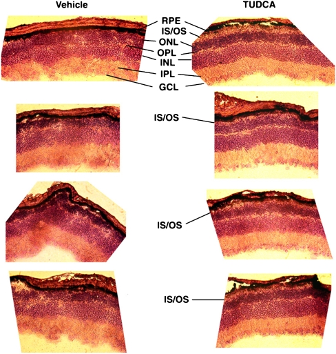
rd1 (rd1) mice were subcutaneously injected with vehicle or TUDCA (500 mg/kg body weight every 3 days). Injections started at postnatal day (P) 9 and continued to P21, at which point animals were killed, and retinal paraffin sections were cut and stained with hematoxylin and eosin (H&E-stained). Vehicle-treated retinas showed the expected near-total loss of ONL cells. Conversely, TUDCA-treated retinas had varied, but more organized morphology ranging from very little ONL to thick ONL and in some instances the preservation of what appear to be photoreceptor outer segments. RPE: retinal pigment epithelium; IS/OS: inner segment/outer segment; ONL: outer nuclear layer; OPL: outer plexiform layer; INL: inner nuclear layer; IPL: inner plexiform layer; GCL: ganglion cell layer
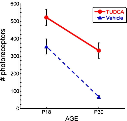
Effect of TUDCA treatment on number of photoreceptor cells in P18 and P30 rd10 mice. rd10 were injected subcutaneously with vehicle or TUDCA (500 mg/kg) every 3 days starting at P6. At P18 and P30 mice were killed, and retinal paraffin sections were prepared and H&E-stained. Outer nuclear layer nuclei were counted in two regions per section and were assumed to reflect number of photoreceptor cells. Each region spanned 400 µm starting from a point 400 µm from either side of the optic nerve. TUDCA treatment significantly preserved the number of photoreceptors at both postnatal days. Of note is that TUDCA treatment delayed the loss of photoreceptor cells by 12 days over the course of the degeneration to P30
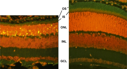
Effect of TUDCA treatment on retinal morphology and TUNEL in P18 rd10 mice. Fluorescence microscopy using a B-2A emission fluorescence filter allows observation of the preservation of photoreceptor inner segments (IS) and outer segments (OS) present in TUDCA- (right) versus vehicle-treated retina sections (left). TUNEL-positive nuclei (green/yellow signal) are seen to be abundant in vehicle-treated sections, but rare retinas from TUDCA-treated mice. TUDCA treatment provided significant preservation of photoreceptor nuclei number in the outer nuclear layer (ONL). Treatment had no discernable effect on the inner nuclear layer (INL) or ganglion cell layer (GCL). Image reprinted with permission from Ref. [81]
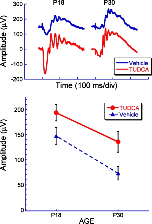
Effect of TUDCA treatment on electroretinograms of rd10 mice at P18 and P30. Representative dark-adapted ERG waveforms (top) and average b-wave amplitude (bottom) to a bright flash (2.1 log cd s/m2) at P18 and P30 in TUDCA and vehicle-treated rd10 mice. Note that TUDCA treatment delayed the loss of retinal function by about 12 days, or about 35%, over the course of the degeneration period (e.g., P30 TUDCA-treated amplitudes are similar to P18 vehicle-treated amplitudes)

Effect of TUDCA on caspase-3 activation in rd10 mouse retina. Paraffin-embedded rd10 mouse retina sections from mice treated with vehicle or TUDCA were assayed for immunoreactivity to activated caspase-3 and observed by confocal microscopy. There was significantly more immunoreactivity (yellow signal) in vehicle-treated sections than in TUDCA-treated sections. Image reprinted with permission from Ref. [81]
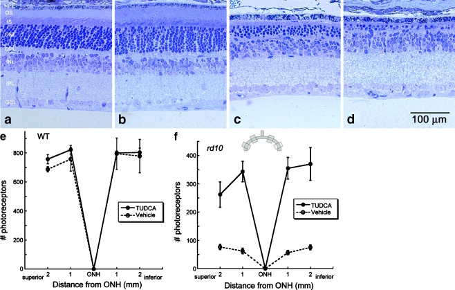
Effect of TUDCA treatment on retinal morphology in rd10 and wild-type mice at P30. Retinal micrographs from P30 wild-type C57BL/6 mice treated with TUDCA (a) or vehicle (b) and from rd10 mice treated with TUDCA (c) or vehicle (d). TUDCA treatment has no effect on wild-type retinal morphology. In rd10 mice, the photoreceptor layer has been reduced to approximately one row of nuclei (d), but TUDCA treatment preserved three to four rows of nuclei (c). c and d Clear differences in the thicknesses of the ONL, outer segments (OS), and inner segments (IS), with TUDCA treatment demonstrating a delay of retinal degeneration. e and f Plots of the total number of photoreceptors at each retinal location from wild type and rd10 mice with reference to the optic nerve head (ONH). The inset is a diagram of the retina and optic nerve and the areas from which cells numbers were sampled. The TUDCA-treated mice have significantly more photoreceptors across all areas sampled than vehicle-treated mice. Image reprinted from Ref. [82]
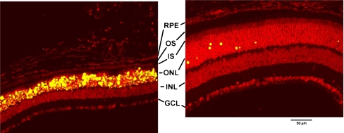
Effect of TUDCA on LIRD mouse retina morphology and apoptosis: 24-h post-light exposure. Mice were subcutaneously injected with either vehicle or TUDCA (500 mg/kg), exposed to 10,000 lx of white light for 7 h, then returned to maintenance lighting conditions. Mice were killed 24 h later, and paraffin retina sections were prepared and assayed for fluorescent TUNEL by confocal microscopy. Representative composite confocal micrographs for vehicle (left) versus TUDCA (right) treatments are shown. Bright-light-exposure-induced massive apoptosis (yellow signal) and morphological damage in photoreceptor cells and retinal outer nuclear layer of vehicle- (left) but not TUDCA-treated eyes (right). Image reprinted with permission from Ref. [81]
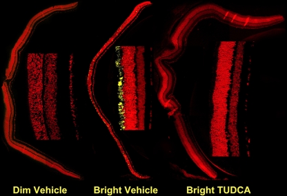
Effect of TUDCA on LIRD mouse retina caspase-3 activation. Mice were subcutaneously injected with either vehicle or TUDCA (500 mg/kg), exposed to 200 (dim) or 10,000 lx (bright) of white light for 7 h, then returned to maintenance lighting conditions. Mice were killed 24 h later, and paraffin retina sections were prepared and probed for activated caspase 3 immunoreactivity by fluorescent confocal microscopy. Representative composite confocal micrographs for vehicle dim-light-exposed vehicle-injected (left), bright-light-exposed vehicle-injected, and bright-light exposed TUDCA-injected (right) treatments are shown. There was significantly more immunoreactivity (yellow signal) in sections of bright-light-exposed vehicle-treated mice than in bright-light-exposed TUDCA-treated mice or dim-light-exposed mice. Image reprinted with permission from Ref. [81]
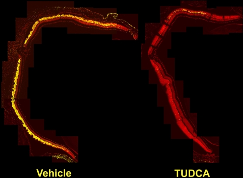
Effect of TUDCA on LIRD mouse retina morphology and apoptosis: 15 days post-light exposure. Mice were subcutaneously injected with either vehicle or TUDCA (500 mg/kg), exposed to 10,000 lx of white light for 7 h, then returned to maintenance lighting conditions. Fifteen days after light exposure, mice were killed, and paraffin retina sections were prepared and assayed for fluorescent TUNEL by confocal microscopy. Bright-light-exposure-induced massive apoptosis and morphological damage in retinal photoreceptors of vehicle-treated (left) but not TUDCA-treated eyes (right). Of particular note in the vehicle-treated sample is the thinning of the outer nuclear layer (ONL) and the nearly complete loss of inner segments (IS) and outer segments (OS) of the photoreceptors. Additionally, nearly all the photoreceptor nuclei are TUNEL positive. Conversely, TUDCA-treated samples showed intact photoreceptors, thick ONL, and very few TUNEL-positive photoreceptor cells. Treatment had no discernable effect on the inner nuclear layer (INL), ganglion cell layer (GCL), or retinal pigment epithelium (RPE). Image reprinted with permission from Ref. [81]
References
-
- Cidian Z. Dictionary of traditional Chinese medicine. Shanghai: Shanghai Science and Technology Press; 2004.
-
- Lee Y-J. The use of bear bile as medicine versus tonic. In: Williamson DF and Phipps MJ, editors. Proceedings of the Third International Symposium on the Trade in Bear Parts. Seoul: TRAFFIC East Asia; 1999. p. 122–126
-
- Gabriel GG.A bitter medicine: the use of bear bile in China. In: Williamson DF and Phipps MJ, editors. Proceedings of the Third International Symposium on the Trade in Bear Parts. Seoul: TRAFFIC East Asia; 1999. p. 116–120.
-
- Hanson DG. Poaching: Oriental demand for undgam and paws decimates California bears. Audubon. 1983;85:127–128.
Grants and funding
LinkOut - more resources
Full Text Sources
Other Literature Sources