. 2009 Dec 31;4(12):e8443.
doi: 10.1371/journal.pone.0008443.

Functional and transcriptional characterization of human embryonic stem cell-derived endothelial cells for treatment of myocardial infarction

Affiliations

Functional and transcriptional characterization of human embryonic stem cell-derived endothelial cells for treatment of myocardial infarction

Zongjin Li et al. PLoS One. .

Abstract

Background: Differentiation of human embryonic stem cells into endothelial cells (hESC-ECs) has the potential to provide an unlimited source of cells for novel transplantation therapies of ischemic diseases by supporting angiogenesis and vasculogenesis. However, the endothelial differentiation efficiency of the conventional embryoid body (EB) method is low while the 2-dimensional method of co-culturing with mouse embryonic fibroblasts (MEFs) require animal product, both of which can limit the future clinical application of hESC-ECs. Moreover, to fully understand the beneficial effects of stem cell therapy, investigators must be able to track the functional biology and physiology of transplanted cells in living subjects over time.

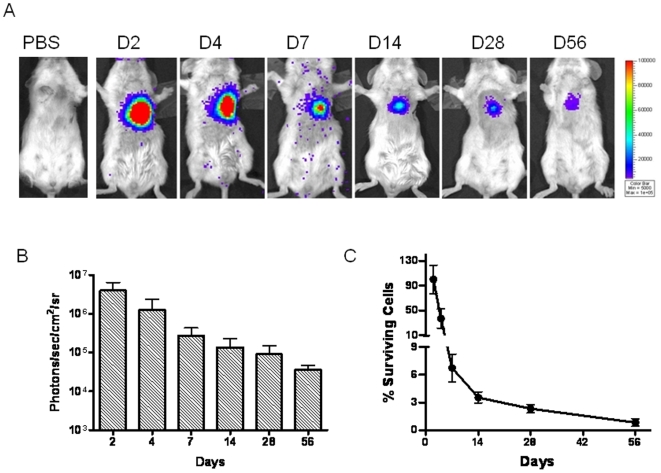
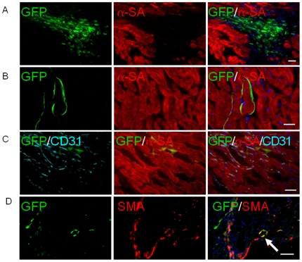
Methodology: In this study, we developed an extracellular matrix (ECM) culture system for increasing endothelial differentiation and free from contaminating animal cells. We investigated the transcriptional changes that occur during endothelial differentiation of hESCs using whole genome microarray, and compared to human umbilical vein endothelial cells (HUVECs). We also showed functional vascular formation by hESC-ECs in a mouse dorsal window model. Moreover, our study is the first so far to transplant hESC-ECs in a myocardial infarction model and monitor cell fate using molecular imaging methods.

Conclusion: Taken together, we report a more efficient method for derivation of hESC-ECs that express appropriate patterns of endothelial genes, form functional vessels in vivo, and improve cardiac function. These studies suggest that hESC-ECs may provide a novel therapy for ischemic heart disease in the future.


Conflict of interest statement

Competing Interests: The authors have declared that no competing interests exist.

Figures

Figure 1
Figure 1. Specification of hESC differentiation into endothelial lineage.
(A) An outline of the protocol used for the differentiation of hESCs to the endothelial lineage. Undifferentiated hESCs were grown to 60%–70% confluence on Matrigel and subcultured in low attachment dish with differentiation medium supplement VEGF and bFGF, shown as day 0. At day 15, EBs in collagen were collected and digested and CD31+/CD144+ cells were isolated by FACS and then sub-cultured in EGM-2 medium with 5% Knockout SRTM to expand and induce endothelial maturation. (B) Whole-mount immunochemistry of day-12 EB. Areas of CD31 (red) and CD144 (green) expression within EBs are organized in elongated clusters and channels. Cell nuclei stained with DAPI (blue). Scale bars = 100 µm. (C) Endothelial differentiation of sprouting EBs in collagen. Representative sprouting EBs after 3 days of culturing in collagen matrix (upper panel, left). Whole-mount immunostaining shows CD31 sprouting with channel-like vessel structures and CD31 cell clumps (upper panel). These sproutings are also CD31 and CD144 double positive. Scale bars = 20 µm (top panel, right one) and 100 µm (others). (D) Kinetic expression of CD31 and CD144 during the two-step hESC differentiation procedures. CD31/CD144 expression was triggered and increased from 1–3% to 10–15% after EBs were subcultured in collagen. hESC-ECs were enriched by isolation CD31+/CD144+ cells after FACS (right panel). Experiments were performed in triplicates.
Figure 2
Figure 2. In vitro characterization of hESC-ECs.
(A) During hESC differentiation and endothelial isolation, FACS analysis shows CD31 and CD144 increase significantly. (B) Morphogenesis shows the cells possess cobblestone-like morphology. Histology shows CD31 and CD144 are expressed on cell membranes, and vWF in the cytoplasm. Scale bars = 20 µm (left), 10 µm (middle two), 5 µm (right). (C) Similar to HUVECs, hESC-ECs also can uptake ac-DiI-LDL and form tube-like structures on Matrigel. Scale bars = 20 µm.
Figure 3
Figure 3. Major themes in gene expression profiles at each stage of differentiation.
(A) hESCs express high levels of pluripotency-associated genes including Oct4, Sox2, NANOG, Lefty, and DNMT. At the EB stage, the cells express high levels of mesodermal master regulators such as Tbx2, and BMP4 as well as very enriched levels of endothelial specific master regulators including EPAS1, TIE2, KDR, and EFNA. This population also expresses genes from other cell layers, and many developmental genes from Wnt and homeobox families. hESC-ECs downregulate early mesodermal genes and express more endothelial specific genes, while HUVECs have the highest levels of mature endothelial gene expression with very few other developmental lineages represented. (B) Principal Components Analysis (PCA) shows that replicate experiments of each cell type are very similar while differentiation groups separate significantly along components 1 and 2. (C) Hierarchical Clustering Analysis - Cells from each developmental stage cluster relatively close to each other, with the most distance between hESCs and HUVECs. (D) K-means clustering analysis identifies major trends in gene expression across the time course.
Figure 4
Figure 4. Quantitative PCR confirmation of endothelial gene expression in hESC, EB, EB sprouting, hESC-EC, and HUVEC.
(A) Scatter plots of the endothelial related gene-expression were compared between hESC vs EB, hESC vs EB sprouting, and EB vs sprouting. Endothelial related genes expression was analyzed by Human Endothelial Cell Biology RT2 Profiler PCR Array. The array includes 84 genes related to endothelial cell biology. The lines indicate the diagonal and 4-fold changes between the two samples. (B) Kinetic expression of selected endothelial genes among hESC, hEB, hEB sprouting, hESC-EC, and HUVEC. Compared to HUVEC, hESC-ECs express abundant endothelial gene except adult endothelial gene vWF. *P<0.05; #P<0.01 compared with HUVECs. (C) After EB embedded into collagen, endothelial differentiation was triggered. Note endothelial activation related gene expression increased swiftly regardless of the level at the beginning.
Figure 5
Figure 5. Demonstration of functional vessels in vivo using Matrigel plug and dorsal window chamber.
(A) Matrigel plug with hESC-ECs were implanted subcutaneous injection in the dorsal region of 8-week-old SCID mice. (I) HE stain of Matrigel plug. Some of these microvessels have mouse blood cells in their lumen. (II–IV) 14-day Matrigel plugs were stained with anti-human CD31 (red) and anti-GFP (green) antibodies, showing microvessels that are immunoreactive with these human-specific antibodies. Scale bars = 20 µm. (B) Dorsal window chamber model in SCID mice. GFP+ hESC-ECs were cultured in a mix of collagen and Matrigel for 1 day (left, upper panel), and implanted into dorsal windows in SCID mice (middle, upper panel). Images were taken at day 2, 14 and 21 after implantation. After 14 days, Angiosense 680 was injected by tail vein to highlight perfused vessels within the dorsal window. Green, hESC-ECs expressing GFP; red, functional blood vessels with contrast enhanced by Angiosense 680. Scale bars = 20 mm (middle, upper panel), 20 µm (others).
Figure 6
Figure 6. Molecular imaging of hESC-EC fate after transplantation.
(A) A representative animal injected with 1×10⁶ hESC-ECs shows significant bioluminescence activity at day 2, which decreases progressively over the following 8 weeks. A representative control animal injected with PBS shows no imaging signals as expected. (B) Detailed quantitative analysis of signals from all animals (n = 28) transplanted with hESC-ECs (signal activity is expressed as photons/sec/cm2/sr). (C) Donor cell survival plotted as % signal activity from day 2 to week 8.
Figure 7
Figure 7. Confirmation of engrafted hESC-ECs by immunofluorescence.
(A) Fate of hESC-ECs within the recipient myocardium 4 days after injection showed clump formation. (B, C, D) At day 28, transplanted hESC-ECs differentiate into vasculature and smooth muscle cells (arrow), and integrate with host myocardium as confirmed by GFP, mouse CD31, α-sarcomeric actin (α-SA) and smooth muscle actin (SMA) co-staining. However, this population became significantly rare compared to day 4 due to donor cell death. Scale bar = 20 µm.

Similar articles

Cited by

References

    1. Wollert KC, Drexler H. Clinical applications of stem cells for the heart. Circ Res. 2005;96:151–163. - PubMed
    1. Laflamme MA, Murry CE. Regenerating the heart. Nat Biotechnol. 2005;23:845–856. - PubMed
    1. Huang PP, Yang XF, Li SZ, Wen JC, Zhang Y, et al. Randomised comparison of G-CSF-mobilized peripheral blood mononuclear cells versus bone marrow-mononuclear cells for the treatment of patients with lower limb arteriosclerosis obliterans. Thromb Haemost. 2007;98:1335–1342. - PubMed
    1. Yang C, Zhang ZH, Li ZJ, Yang RC, Qian GQ, et al. Enhancement of neovascularization with cord blood CD133+ cell-derived endothelial progenitor cell transplantation. Thromb Haemost. 2004;91:1202–1212. - PubMed
    1. Heeschen C, Chang E, Aicher A, Cooke JP. Endothelial progenitor cells participate in nicotine-mediated angiogenesis. J Am Coll Cardiol. 2006;48:2553–2560. - PubMed

Publication types

MeSH terms