. 2009;43(8):1171-1178.
doi: 10.1016/j.asr.2008.10.011.

γ-H2AX as a biomarker of DNA damage induced by ionizing radiation in human peripheral blood lymphocytes and artificial skin

Affiliations

γ-H2AX as a biomarker of DNA damage induced by ionizing radiation in human peripheral blood lymphocytes and artificial skin

Christophe E Redon et al. Adv Space Res. 2009.

Abstract

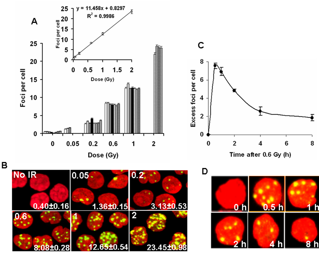
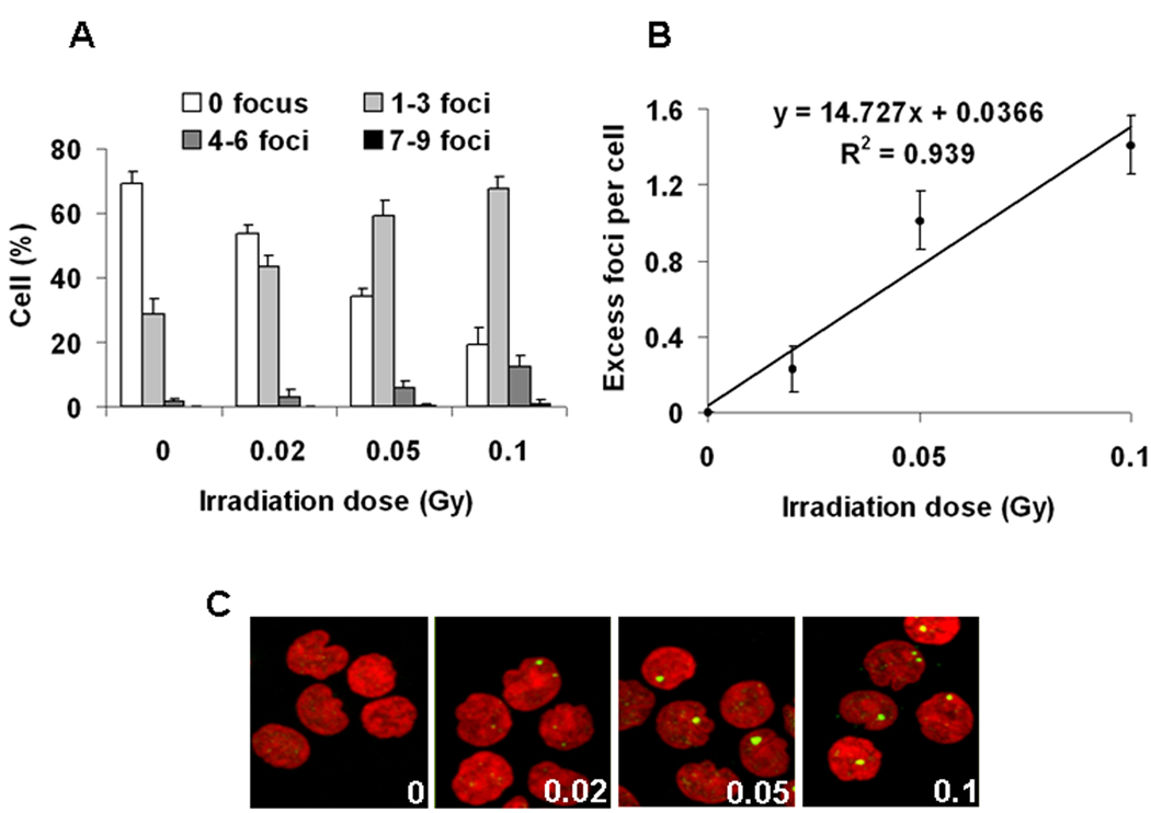
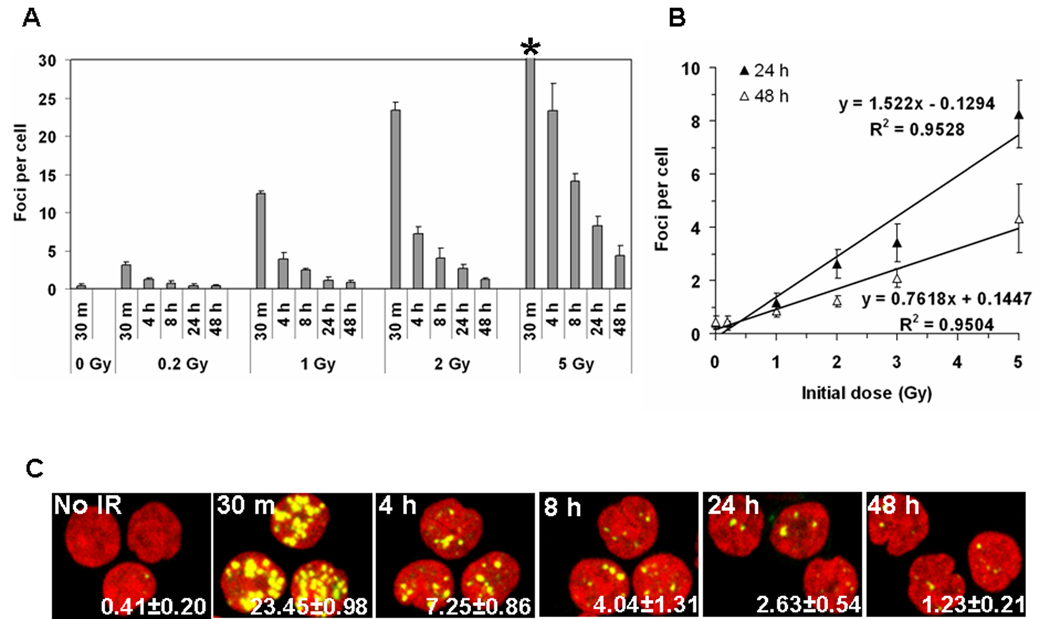
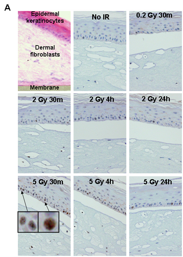
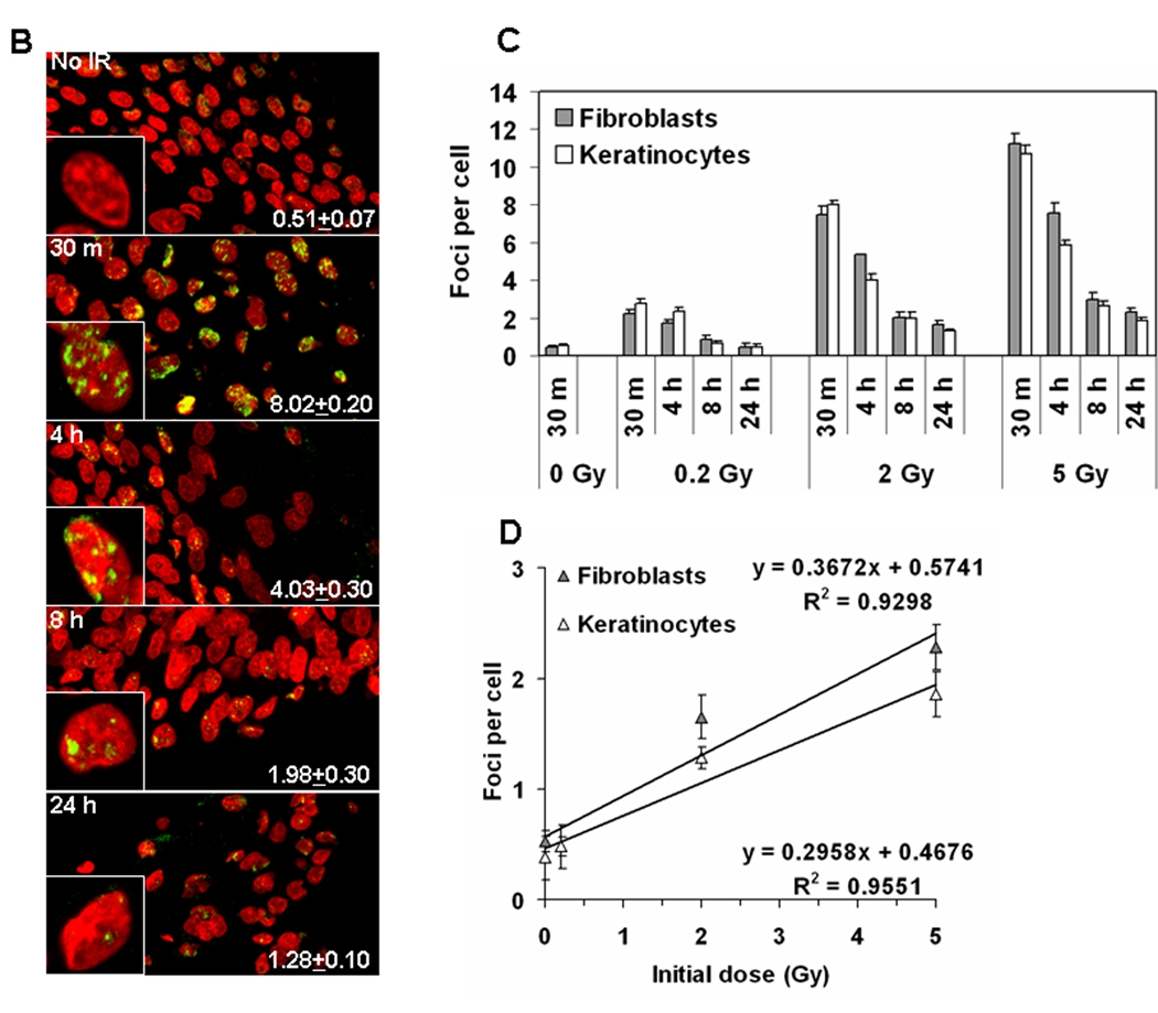
Ionizing radiation (IR) exposure is inevitable in our modern society and can lead to a variety of deleterious effects including cancer and birth defects. A reliable, reproducible and sensitive assessment of exposure to IR and the individual response to that exposure would provide much needed information for the optimal treatment of each donor examined. We have developed a diagnostic test for IR exposure based on detection of the phosphorylated form of variant histone H2AX (γ-H2AX), which occurs specifically at sites of DNA double-strand breaks (DSBs). The cell responds to a nascent DSB through the phosphorylation of thousands of H2AX molecules flanking the damaged site. This highly amplified response can be visualized as a γ-H2AX focus in the chromatin that can be detected in situ with the appropriate antibody. Here we assess the usability of γ-H2AX focus formation as a possible biodosimeter for human exposure to IR using peripheral blood lymphocytes irradiated ex vivo and three-dimensional artificial models of human skin biopsies. In both systems, the tissues were exposed to 0.2-5 Gy, doses of IR that might be realistically encountered in various scenarios such as cancer radiotherapies or accidental exposure to radiation. Since the γ-H2AX response is maximal 30 minutes after exposure and declines over a period of hours as the cells repair the damage, we examined the time limitations of the useful detectibility of γ-H2AX foci. We report that a linear response proportional to the initial radiation dose was obtained 48 hours and 24 hours after exposure in blood samples and skin cells respectively. Thus, detection of γ-H2AX formation to monitor DNA damage in minimally invasive blood and skin tests could be useful tools to determine radiation dose exposure and analyze its effects on humans.


Figures

Figure 1
Figure 1
DNA damage in peripheral blood lymphocytes exposed to IR ex vivo. (A) (Main graph) Incidence of γ-H2AX foci in blood taken from eight individual donors and irradiated ex vivo. Lymphocytes were purified 30 minutes post-IR, stained for γ-H2AX detection and then γ-H2AX foci were counted. Each donor is noted with a differently patterned bar. Error bars indicate standard errors. (Inset) Average numbers of γ-H2AX foci per cell were determined. Error bars indicate standard deviations (n=8). (B) Representative images of irradiated lymphocytes used for panel A. The irradiation dose (Gy) is shown in the top left corner and the number of γ-H2AX foci per cell is shown in the lower right corner (average ±standard error). Green, γ-H2AX; red, DNA stained with PI. (C) Kinetics of γ-H2AX focal loss in lymphocytes after 0.6 Gy IR. Blood samples were irradiated with 0.6 Gy and incubated at 37°C. At indicated times lymphocytes were isolated and stained for γ-H2AX detection. Error bars indicate standard deviations (n=3). (D) Representative images of irradiated lymphocytes used for panel C. Green, γ-H2AX; red, DNA stained with propidium iodide (PI).
Figure 2
Figure 2
DNA damage in human peripheral blood lymphocytes exposed ex vivo to low IR doses. (A) Focal distribution in lymphocytes 30 minutes after exposure to 0, 0.02, 0.05 and 0.1 Gy. (B) Average numbers of excess (background subtraction) γ-H2AX foci per cell as determined 30 minutes after 0.02, 0.05 and 0.1 Gy. Error bars indicate standard deviations (n=3). (C) Representative images of irradiated lymphocytes used for panel A. The irradiation dose (Gy) is shown in the lower right corner. Green, γ-H2AX; red, DNA stained with PI.
Figure 3
Figure 3
Kinetics of long-term γ-H2AX focal loss in blood from five individuals after IR (A) The incidence of γ-H2AX foci in lymphocytes irradiated with 0, 0.2, 1.0, 2.0, and 5.0 Gy taken at the noted times post-exposure. (*) The foci 30 minutes after 5 Gy were too numerous to count. (B) The incidence of γ-H2AX foci at 24 hours and 48 hours post-IR exposure is shown. Error bars signify standard deviations (n=5). (C) Representative images of irradiated lymphocytes used for panel A. Time post-IR is shown in the top left corner and the number of γ-H2AX foci per cell is shown in the lower right corner (average±standard error). Green, γ-H2AX; red, DNA stained with PI.
Figure 4
Figure 4
IR-induced DNA damage in artificial human skin. (A) Peroxidase staining in paraffin-embedded tissues. The EpiDermFT is a “full thickness” highly differentiated artificial skin consisting of epidermal keratinocyte and dermal fibroblast layers, corresponding to dermis and epidermis of normal human skin. The tissues were formalin-fixed and paraffin-embedded at various times after radiation exposure, and sectioned perpendicularly to their surface. (Upper left panel) Sections stained with hematoxilin and eosine. The tissue is 300 µm high grown on a 6 mm diameter cellulose membrane. Blue staining indicates cell nuclei while pink staining indicates cytoplasm. (Other panels) γ-H2AX staining in tissues irradiated with 0, 0.2, 2.0, and 5.0 Gy and fixed at various times post-IR. Blue staining is cell nuclei while brown (peroxidase) staining indicates γ-H2AX formation. Two patterns of positive reaction, punctate staining and a whole-nucleus staining (insets), were observed in 2 Gy- and 5 Gy-irradiated samples. At 24 hours post-IR some residual staining was observed. 0.2 Gy-irradiated samples exhibited no or negligible staining. All images are shown at 20 × magnification. (B) Immunostaining of γ-H2AX in frozen tissue sections. Representative images showing the presence of γ-H2AX foci in the keratinocyte EpiDermFT layer irradiated with 0 or 2 Gy. Green, γ-H2AX; red, DNA stained with propidium iodide (PI). Insets show representative single cell images. Average numbers of foci per cell ± standard error are shown in the right bottom corner of the images. (C) Quantitative measure of the incidence of γ-H2AX foci in fibroblasts (grey bars) and keratinocytes (white bars) irradiated with 0, 0.2, 2.0, and 5.0 Gy at various times post-exposure. (D) γ-H2AX focal numbers at 24 hours post-exposure are proportional to the initial dose of IR received in both fibroblasts and keratinocytes. Error bars signify standard errors.
Figure 4
Figure 4
IR-induced DNA damage in artificial human skin. (A) Peroxidase staining in paraffin-embedded tissues. The EpiDermFT is a “full thickness” highly differentiated artificial skin consisting of epidermal keratinocyte and dermal fibroblast layers, corresponding to dermis and epidermis of normal human skin. The tissues were formalin-fixed and paraffin-embedded at various times after radiation exposure, and sectioned perpendicularly to their surface. (Upper left panel) Sections stained with hematoxilin and eosine. The tissue is 300 µm high grown on a 6 mm diameter cellulose membrane. Blue staining indicates cell nuclei while pink staining indicates cytoplasm. (Other panels) γ-H2AX staining in tissues irradiated with 0, 0.2, 2.0, and 5.0 Gy and fixed at various times post-IR. Blue staining is cell nuclei while brown (peroxidase) staining indicates γ-H2AX formation. Two patterns of positive reaction, punctate staining and a whole-nucleus staining (insets), were observed in 2 Gy- and 5 Gy-irradiated samples. At 24 hours post-IR some residual staining was observed. 0.2 Gy-irradiated samples exhibited no or negligible staining. All images are shown at 20 × magnification. (B) Immunostaining of γ-H2AX in frozen tissue sections. Representative images showing the presence of γ-H2AX foci in the keratinocyte EpiDermFT layer irradiated with 0 or 2 Gy. Green, γ-H2AX; red, DNA stained with propidium iodide (PI). Insets show representative single cell images. Average numbers of foci per cell ± standard error are shown in the right bottom corner of the images. (C) Quantitative measure of the incidence of γ-H2AX foci in fibroblasts (grey bars) and keratinocytes (white bars) irradiated with 0, 0.2, 2.0, and 5.0 Gy at various times post-exposure. (D) γ-H2AX focal numbers at 24 hours post-exposure are proportional to the initial dose of IR received in both fibroblasts and keratinocytes. Error bars signify standard errors.

References

    1. Belyakov OV, Mitchell SA, Parikh D, Randers-Pehrson G, Marino SA, Amundson SA, Geard CR, Brenner DJ. Biological effects in unirradiated human tissue induced by radiation damage up to 1 mm away. Proc Natl Acad Sci U S A. 2005;102:14203–14208. - PMC - PubMed
    1. Boelsma E, Gibbs S, Faller C, Ponec M. Characterization and comparison of reconstructed skin models: morphological and immunohistochemical evaluation. Acta Derm Venereol. 2000;80:82–88. - PubMed
    1. Cucinotta FA, Pluth JM, Anderson JA, Harper JV, O'Neill P. Biochemical kinetics model of DSB repair and induction of gamma-H2AX foci by non-homologous end joining. Radiat Res. 2008;169:214–222. - PubMed
    1. Francastel C, Schubeler D, Martin DI, Groudine M. Nuclear compartmentalization and gene activity. Nat Rev Mol Cell Biol. 2000;1:137–143. - PubMed
    1. Goodarzi AA, Noon AT, Deckbar D, Ziv Y, Shiloh Y, Lobrich M, Jeggo PA. ATM signaling facilitates repair of DNA double-strand breaks associated with heterochromatin. Mol Cell. 2008;31:167–177. - PubMed